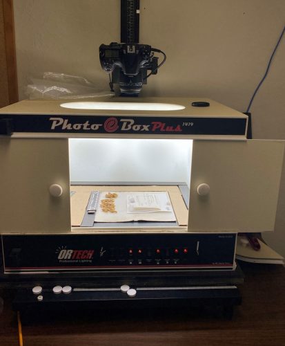
SPECIMEN IMAGING SYSTEM (2022-present)
Digitization of bryophyte and lichen packets requires imaging the actual physical specimen and the label. The actual bryophyte plants or lichen fungi are removed from the packet and placed on a card, with the packet and label adjacent. Then the whole thing is photographed, the organism plus the label.
Our current imaging setup uses a Nikon D180 camera body with a Sigma 50mm 1:2.8 DG Macro lens. The lighting system utilizes a Photo eBox Plus 1419 (which is no longer serviced, so new LED lights will be used once the original eBox lights go out).
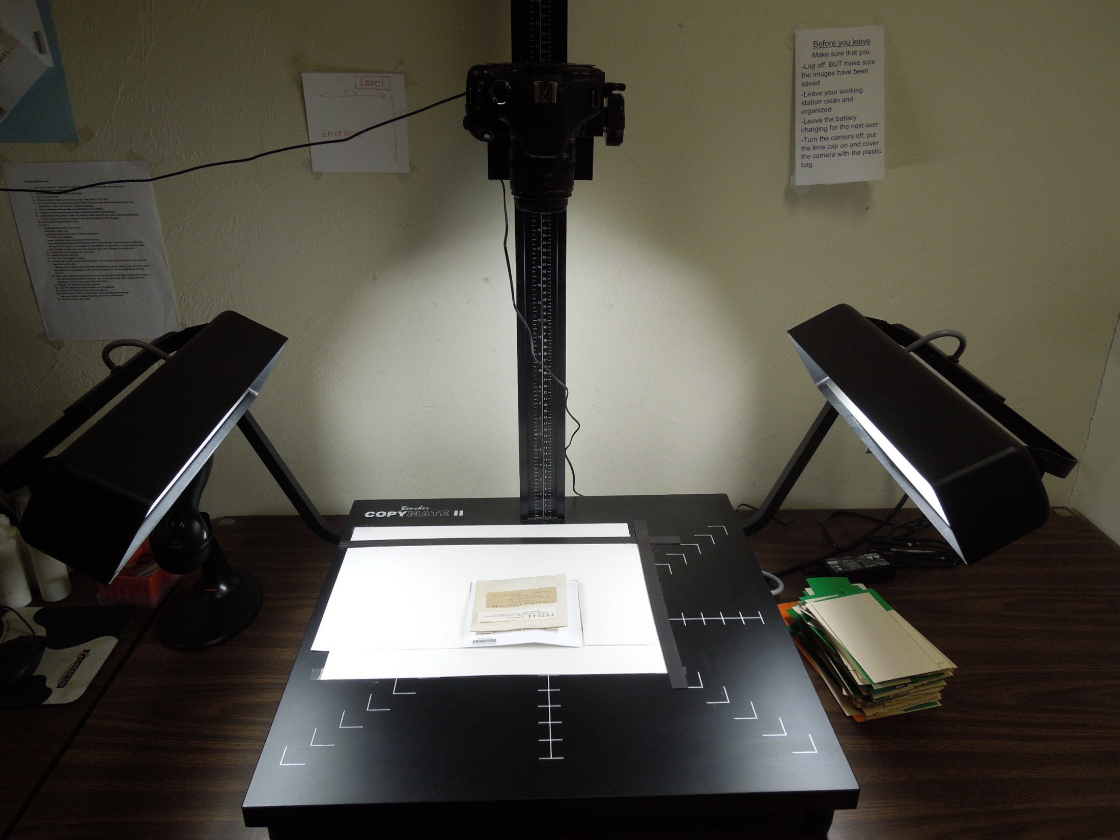
LABEL IMAGING SYSTEM (2012-2021)
For imaging strictly the packet labels, the old imaging setup was composed of a Bencher Copymate III Fluorescent Tabletop Producer with two cool, full spectrum fluorescent lamps that were daylight balanced at 5200°K for color clarity and operated at 12,000 Hz to eliminate flickering. The fluorescent system maintained a cool temperature, and it offered a full spectrum / daylight balanced light. The copystand included an aluminum column that supported a 35 mm Canon EOS T3i digital camera with a Canon EF 50mm f/2.5 compact macro lens, connected to an AC Canon Adapter kit. The system is connected to the PC computer of the project, and we capture the images using the Canon EOS Utility software coupled with the Imaging Workflow Application provided by on the LBCC – Lichens, Bryophytes and Climate Change Web site. In order to capture the image, small earth magnets (7 mm diameter) were used to fix the specimens to the copystand. The magnets were painted white with an industrial grade paint marker.
Imaging of bryophyte/lichen labels averaged from 80-100 per hour. Two images are necessary when there are annotations covering portions of the label. If a group had many annotations the imaging process was considerably slower.