I spent a few days getting some anatomical drawings done. They’re not my first. I have a long history in art. I started learning from my first art teacher, my mom, when I was about 5. Growing up, I’d get in trouble in my art classes for doing my own concepts instead of my assigned drawings. In high school, I was accepted to the Cooper Union Saturday art program, an intense all day drawing regiment, open to New York City high school students, which pushed my artwork to new levels and taught me how to systematically improve my artwork. My senior year I won an entrepreneurship contest and was awarded start up money and a space in the high school’s mini mall to start a small custom art business that I’ve maintained in one form or another over the years, teaching myself to use airbrush, Photoshop, and ZBrush along the way. While at the American Museum of Natural History working on a skull of one of my favorite groups of dinosaurs, ankylosaurs, I became obsessed with reconstructing what I envisioned that animal to look like alive. It took a lot of work, but I successfully completed my reconstruction of the Mongolian armored dinosaur, Minotaurasaurus (recently synonymized with Tarchia) which I was happy to include in my SVP presentation on that specimen.
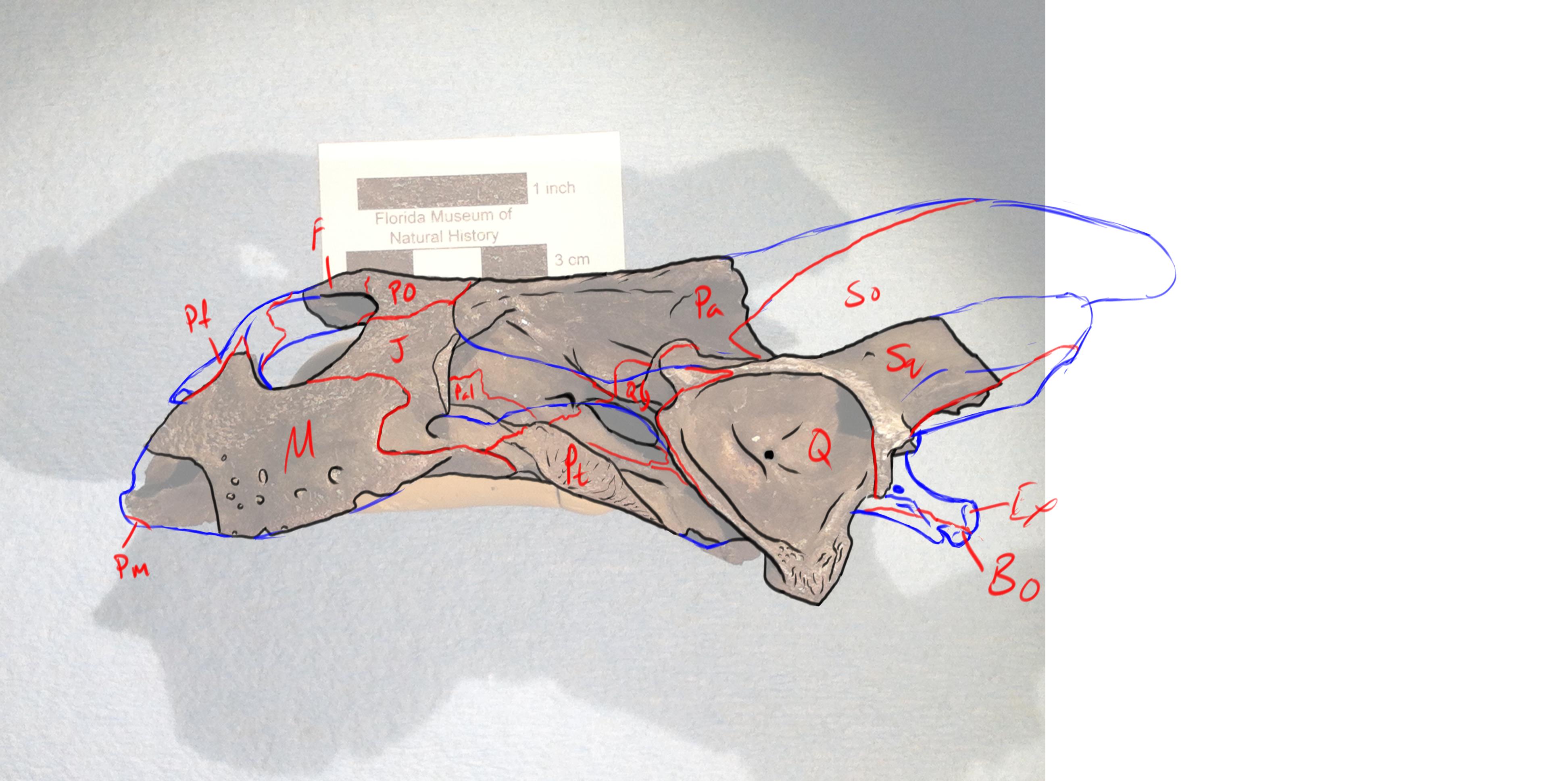
So this week, in an effort to solidify my knowledge of turtle anatomy, I decided to draw a series of scientific illustrations of the fossil skull I am working on, with successive layers of added information baked in. One layer that included the contour line drawing of the skull based on a picture, one including a map of the sutures of the skull, another including the labels of all the bones of the skull and another with a reconstruction that included the missing cranial elements as compared to a modern skull.
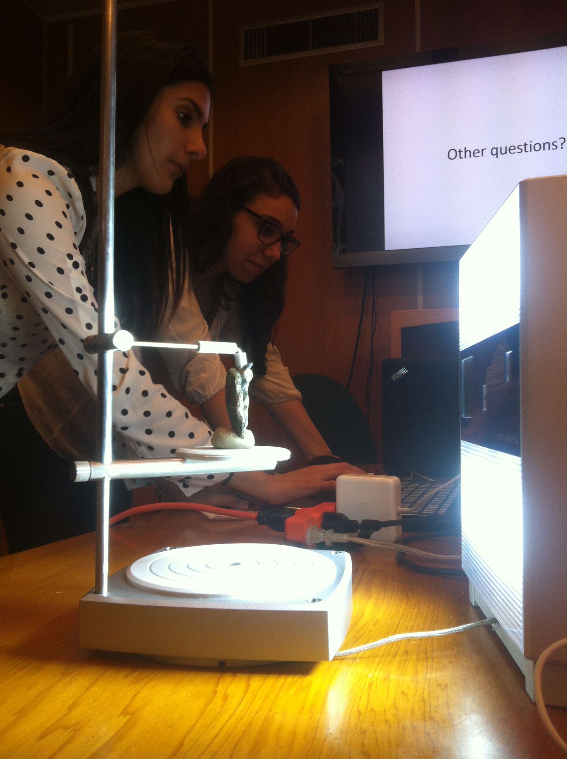
I also got to put my interest in art and technology to work making 3d images of fossils. We are using 2 very different approaches. One uses CT images of the fossil and the other pictures and lasers to generate 3 dimensional images. CT scanning involves taking serial scans through a subject. These images are then taken and, through a series of steps on the computer, joined slice by slice to reconstitute the image of the original object. Surface scanning involves taking pictures of a rotated object and, using a laser to judge distance, stitching the pictures together to get a 3d representation of the contour of the material. Because of the limits of these techniques there are advantages and disadvantages associated with both. Either way it’s still pretty cool being able to create and manipulate a digital replica of your specimen. Trying to troubleshoot some of our work has led to some really interesting discoveries as to what we are and aren’t able to do with the approaches and I’m very interested in finding possible ways around some of these limitations.
While all that is going on, we prepare for our field trip to Panama. We’re off this weekend and eager to play in the dirt. I’m not looking forward to the heat and humidity we will surely encounter but the prospect of finding some great fossils in such a historic location in another country makes it all worth it. The energy here is excited with everyone anxious for the trip. A full account to be provided when we return.