We have a number of active research projects that investigate the structure, function, and evolution of anatomy in amphibians and reptiles. Not only are we interested to document patterns of anatomical diversity, we also seek to understand the processes that generated variation across species.
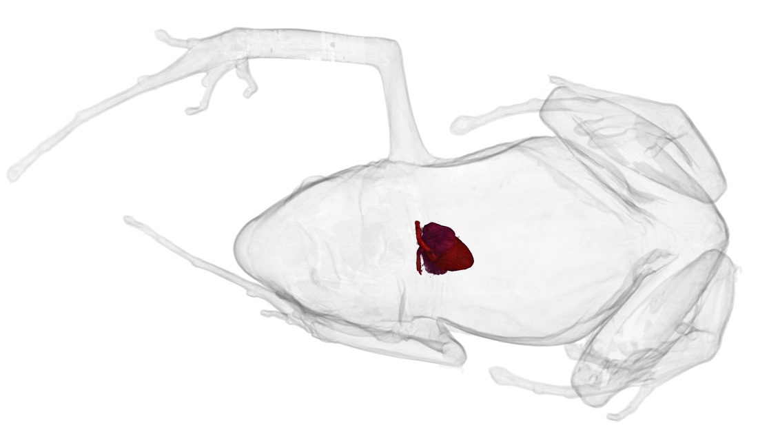
For the most part, this research is driven by high-resolution computed tomography (CT-scanning), which is a powerful method for visualizing, measuring, and disseminating information about anatomy. Because CT-scanning is non-destructive, we can use this technique to study the internal anatomy of rare or delicate museum specimens. Because the resulting data are digital, they can be easily and instantly shared with scientists, students, and the public. We use the CT-scanner based at UF’s Nanoscale Research Facility.
From 2017–2023, we led the oVert (openVertebrate) Thematic Collections Network funded by the US National Science Foundation. This network included participation from 16 museum institutions in the US. We CT-scanned >13,000 vertebrate specimens in US museum collections to provide high-resolution digital anatomy for most vertebrate genera. These data are available via the on-line repository MorphoSource. Read more here in Science and here in BioScience.
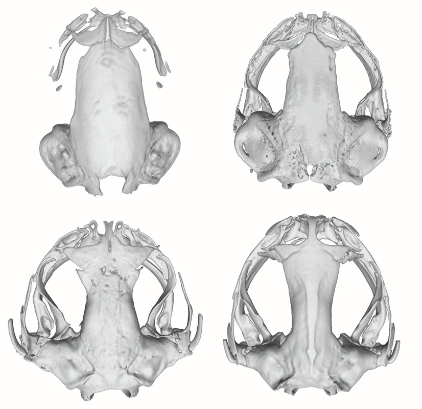
In addition to sharing 3D anatomical data to facilitate research, we are also interested in using these data for education and outreach. We have a diversity of 3D models available on-line at Sketchfab, including examples of all living frog families and vertebrate skull anatomy.