Inner Beauty was on display in the Florida Museum from July 2021 through May 2023. It was so popular, we reimagined it as an online exhibit.
Museum technology is shifting how fish skeletons are processed providing more accessibility for research, education, outreach and global collaboration.
Museum Skeleton Prep
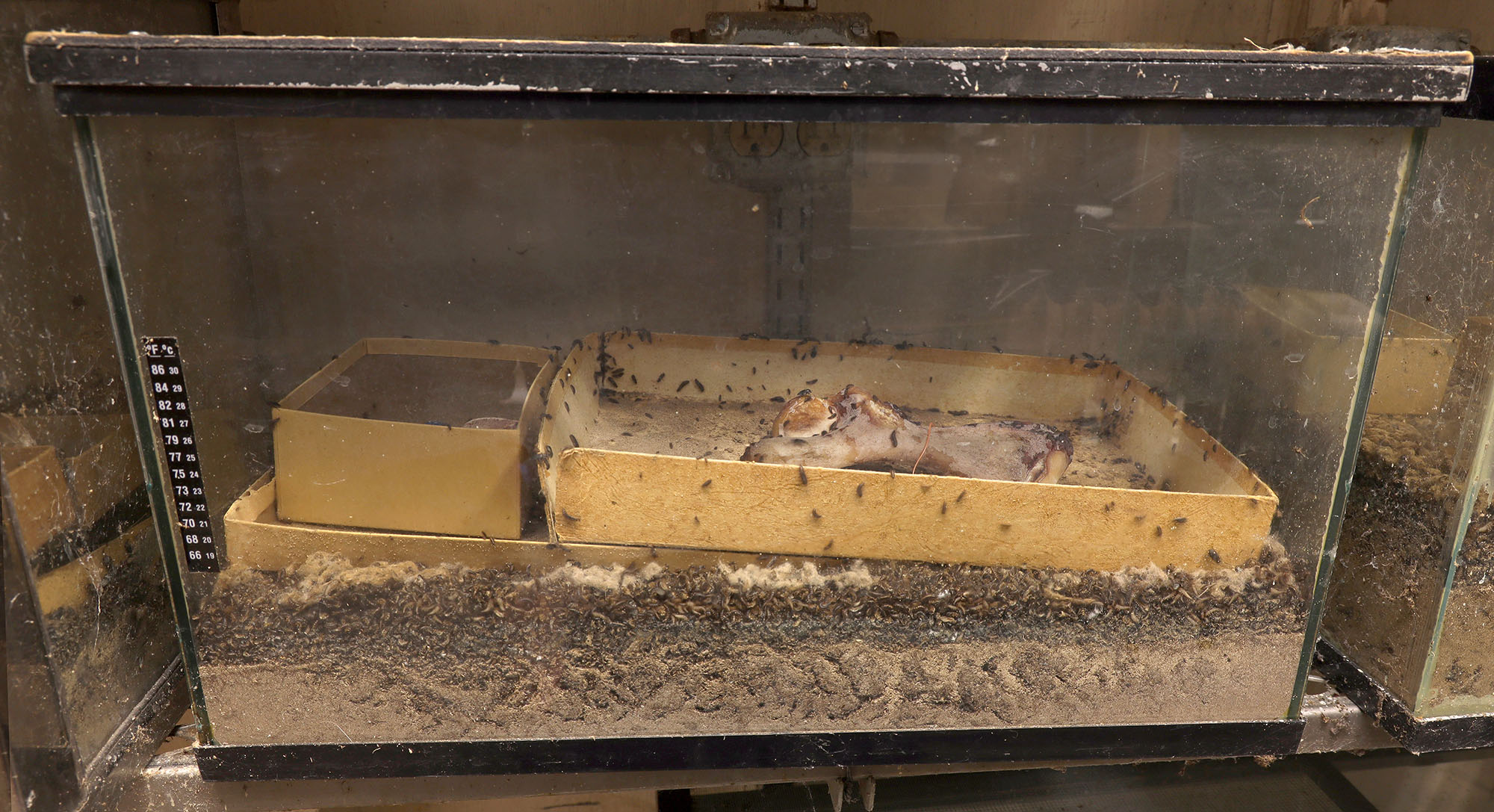
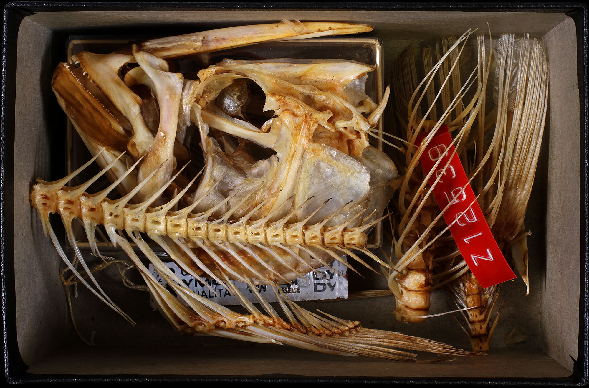
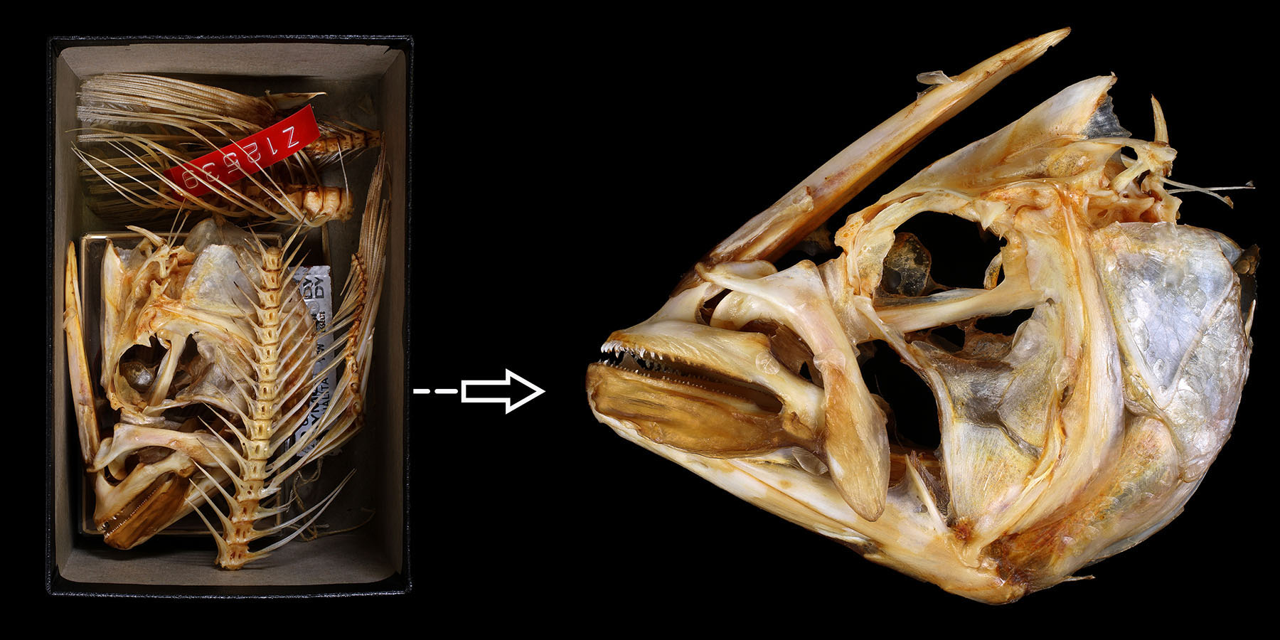
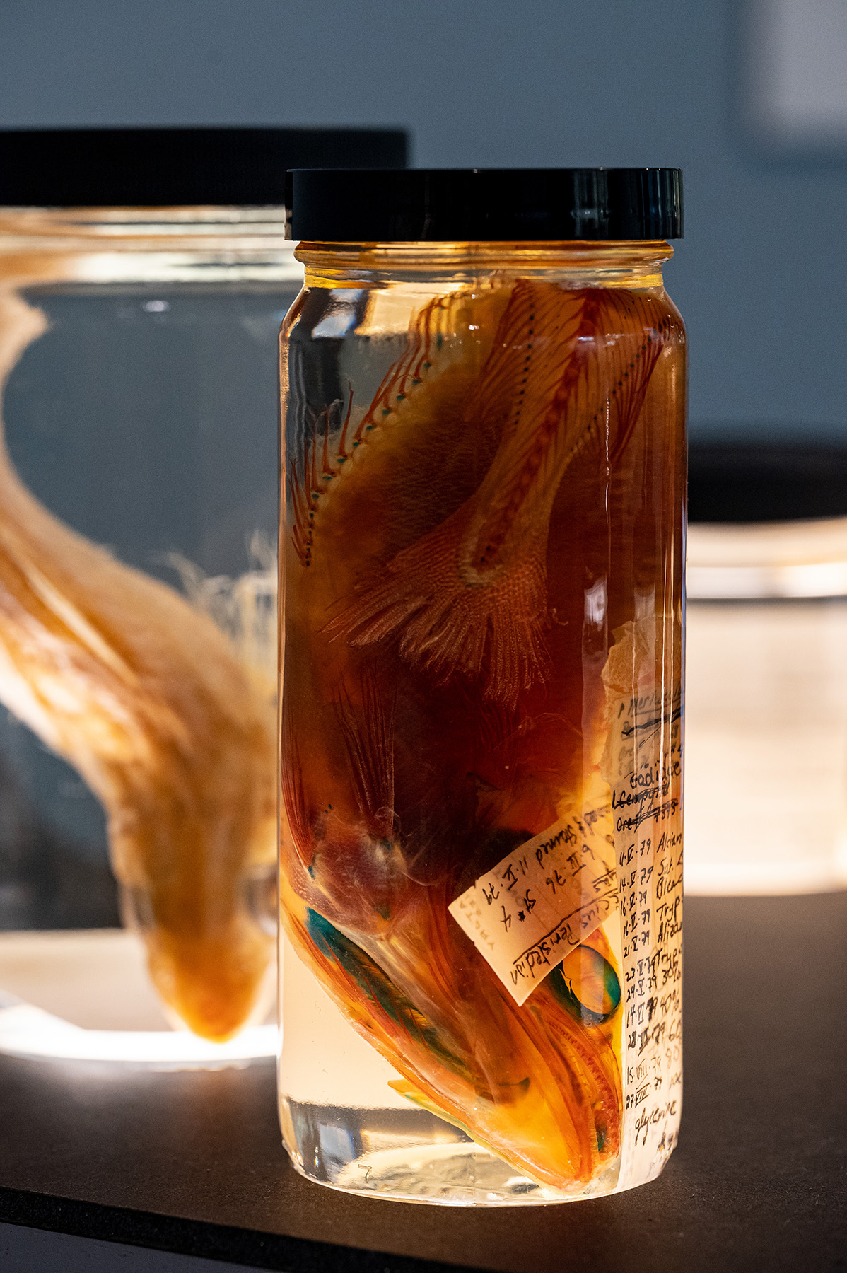
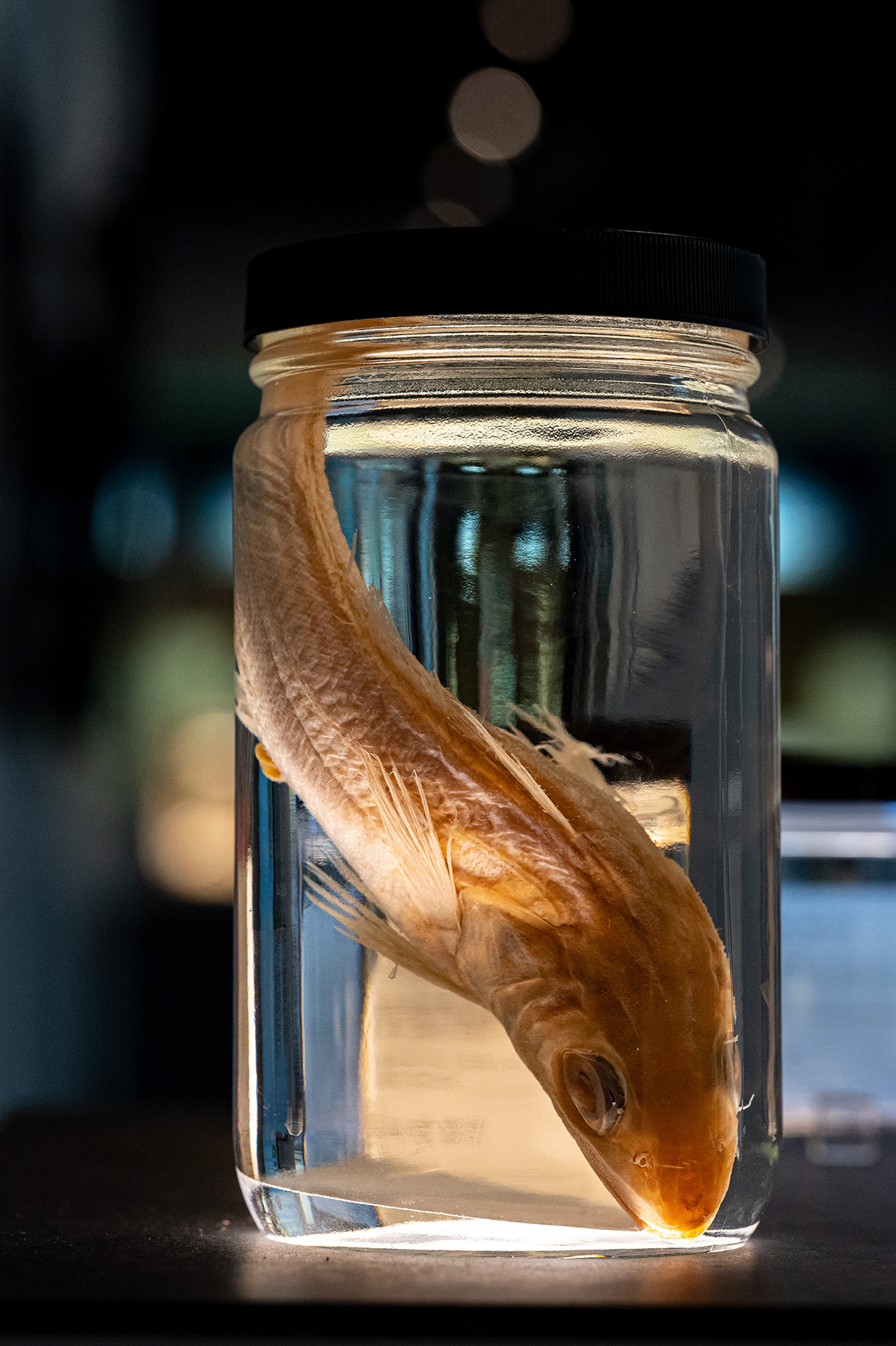
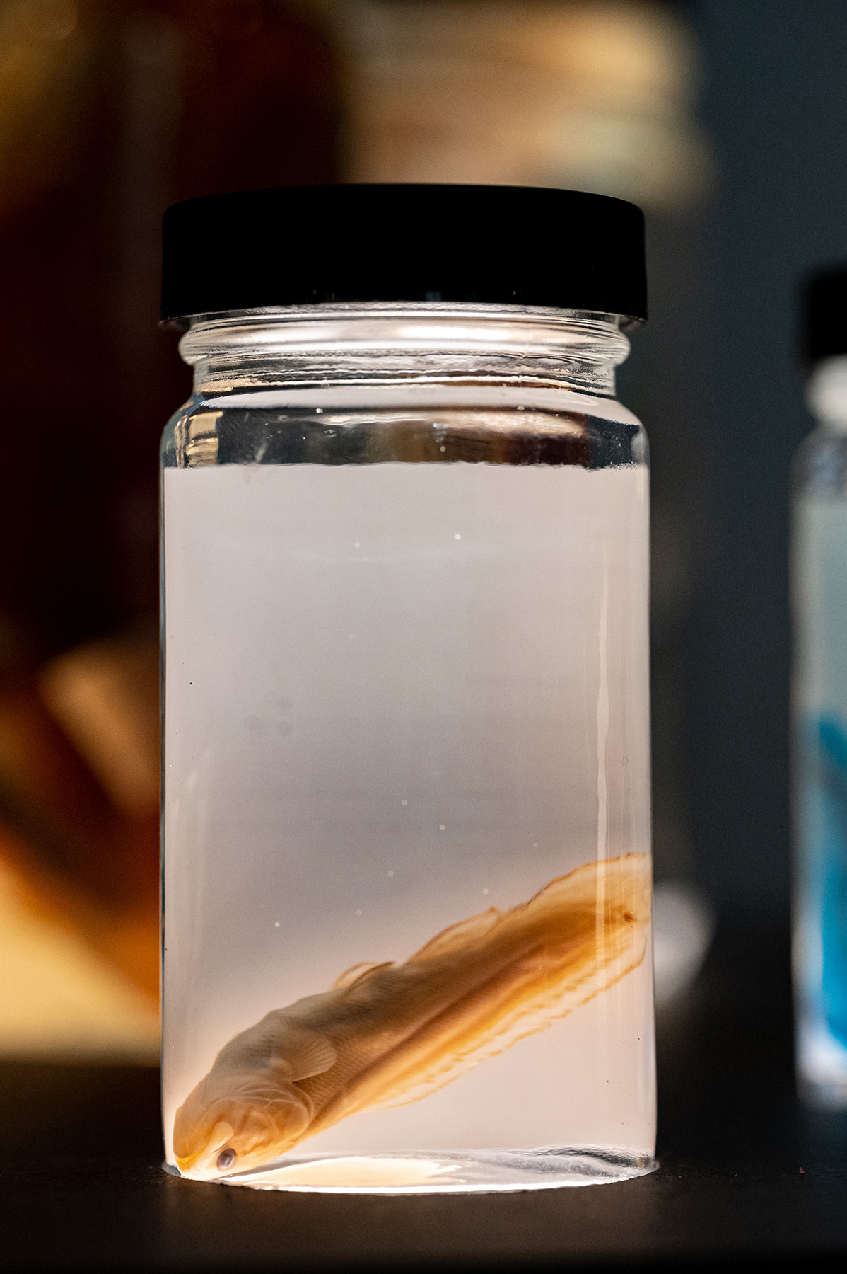
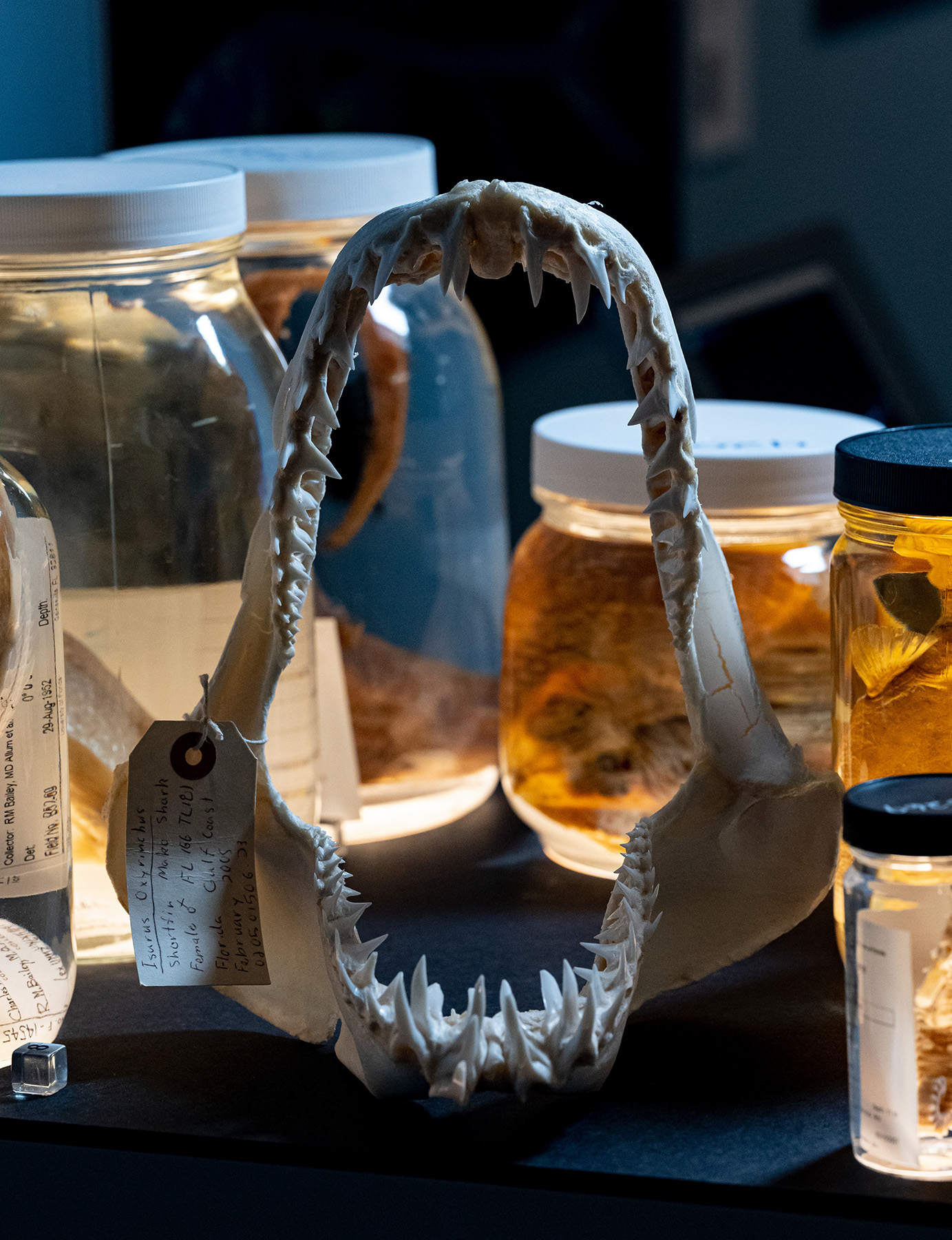
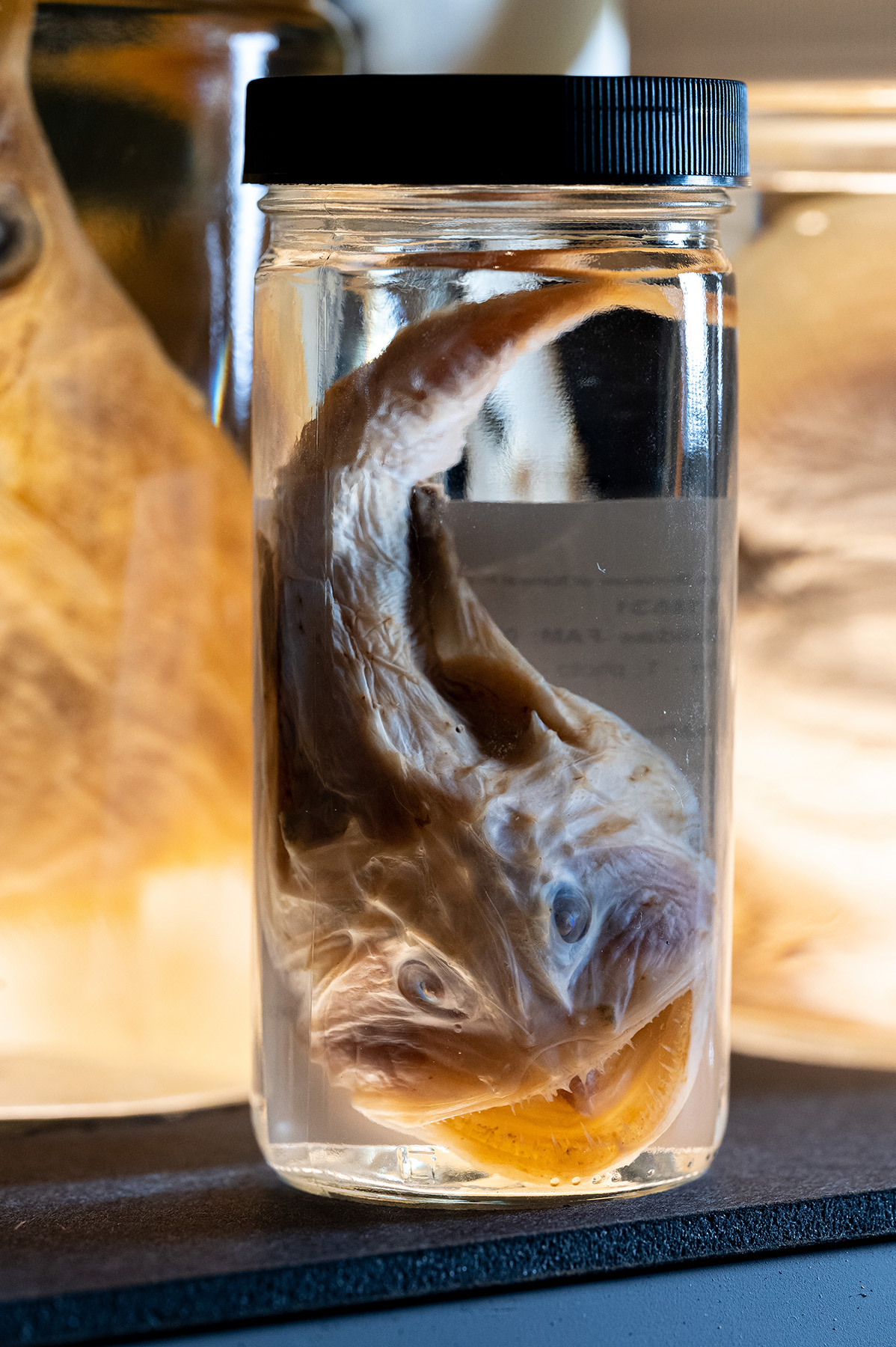
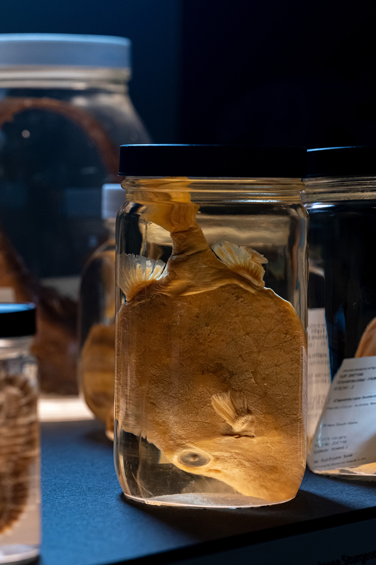
The majority of non-fossilized skeletons at the Florida Museum are processed in-house from preserved museum specimens by using flesh-eating Dermestid Beetle colonies that eat the flesh right off the bones. Specimens are skinned, dissected, dried and then placed in a container with beetles that eat away the dried soft tissue, similar to eating tasty beef jerky. The bones are then disarticulated (separated) and in this process the natural body position is lost. When skeletons of all sizes are being compared, this makes it difficult to study evolutionary relationships and development.
Skeletal Transformation: A Long-term Effect
The skeletonizing method chosen has a direct long-term effect on the specimen. Using either the Dermestid Beetle colony or the clearing and staining method completely transforms the specimen into a skeleton. Important anatomical and biological information such as muscles and diet content are destroyed forever. This poses a challenge for future researchers. These methods cannot be used on rare and irreplaceable specimens. The strength of using CT scanning is that it provides a digital skeleton without altering or damaging the specimen.
Keeping the Bones Together
Ichthyologists (fish biologists) use two main techniques to prepare skeletons from preserved specimens of all shapes and sizes without losing their natural skeletal positions: clearing and staining and computed tomography (CT) scanning.
Clearing and Staining

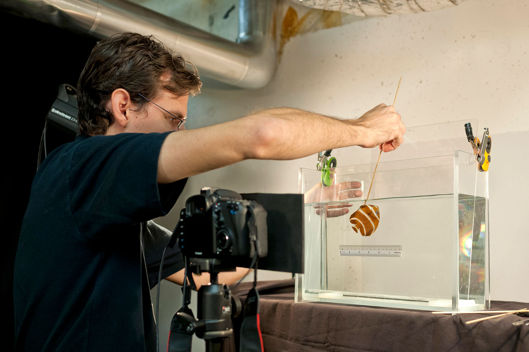
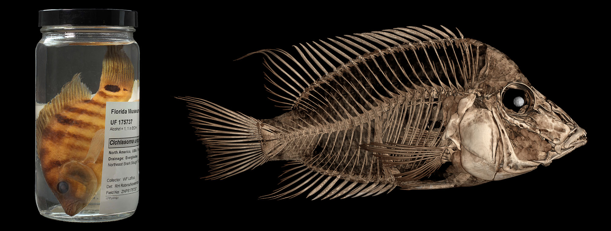
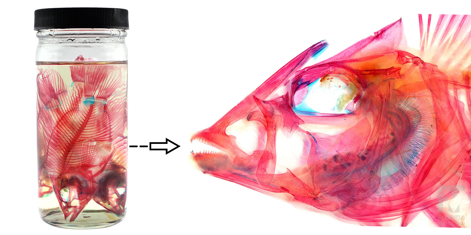
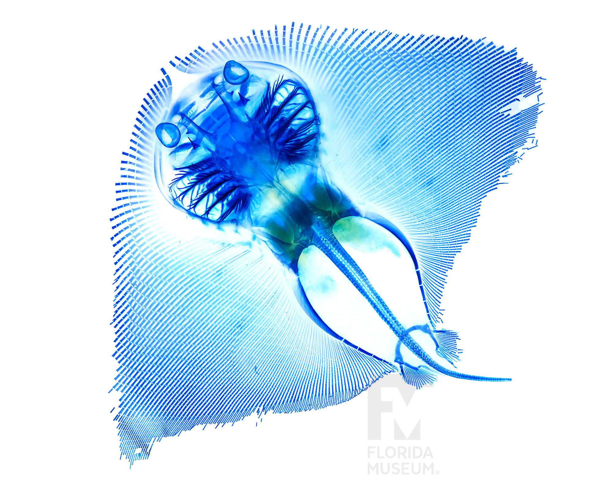
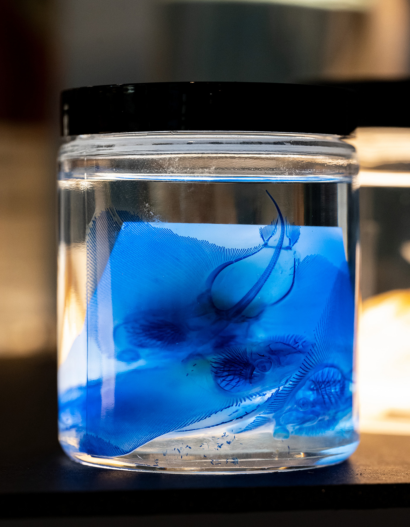
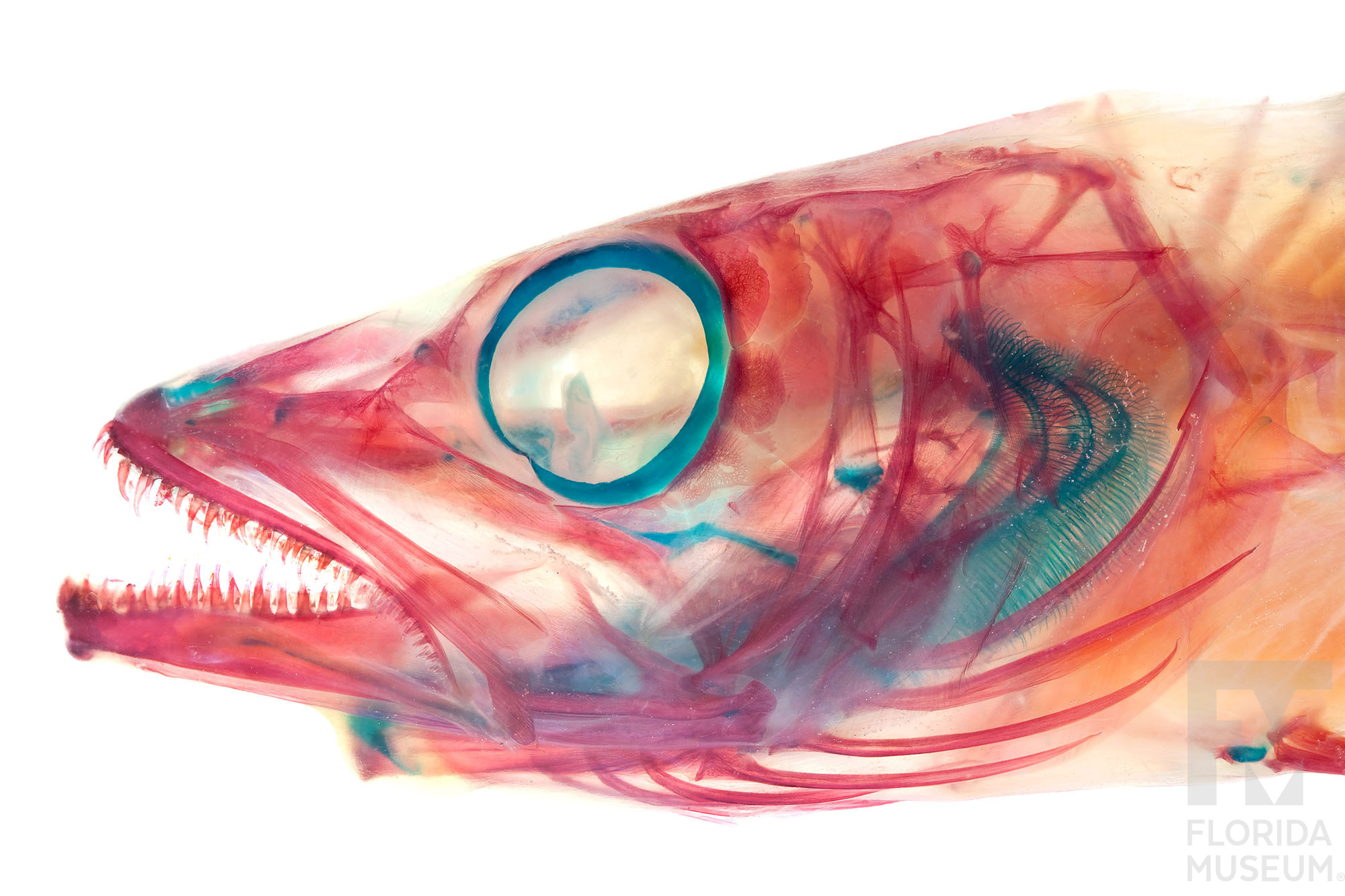
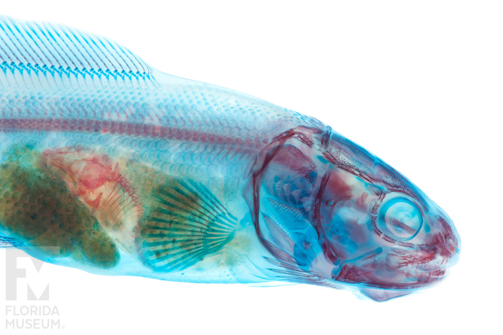
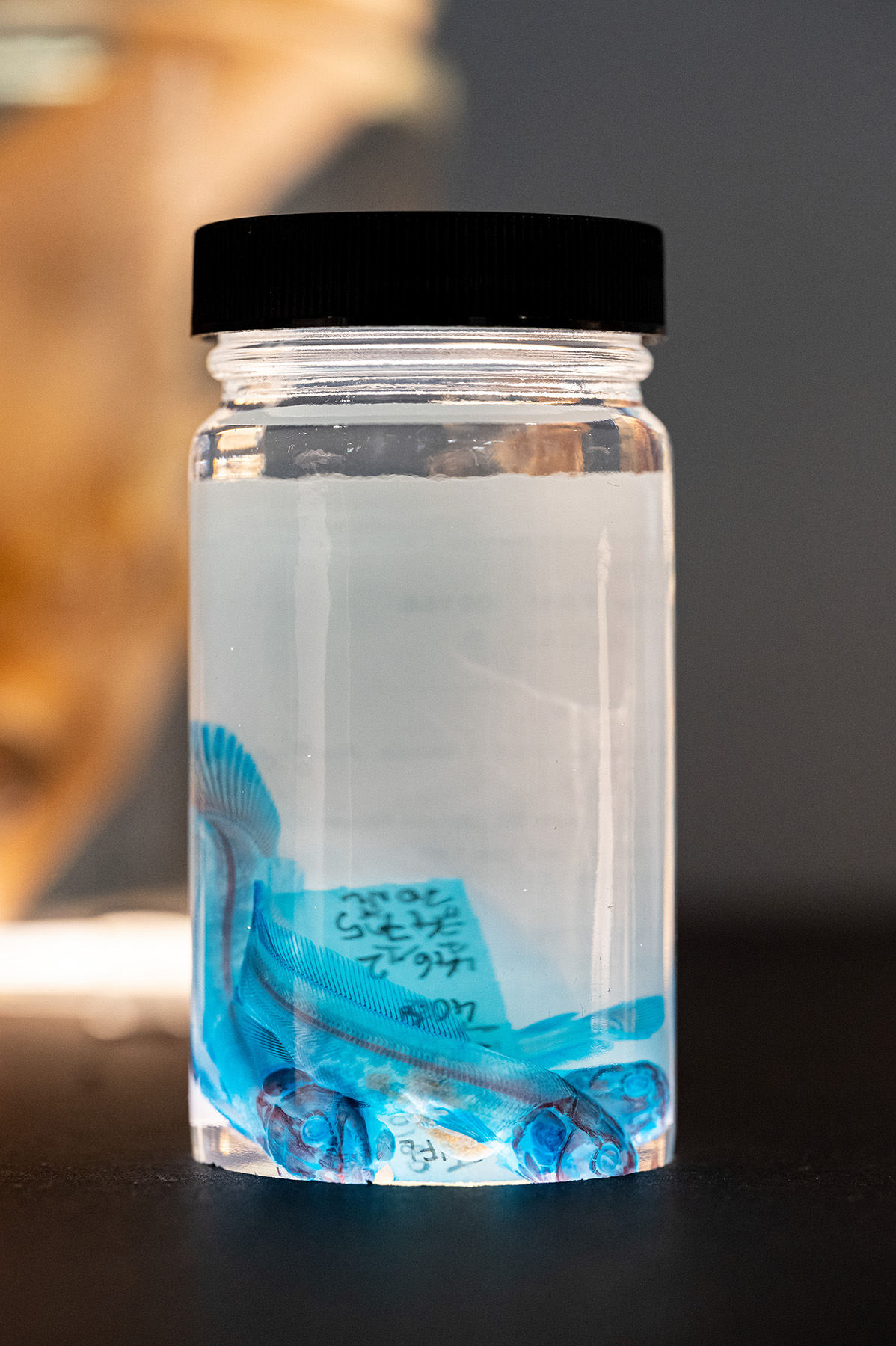
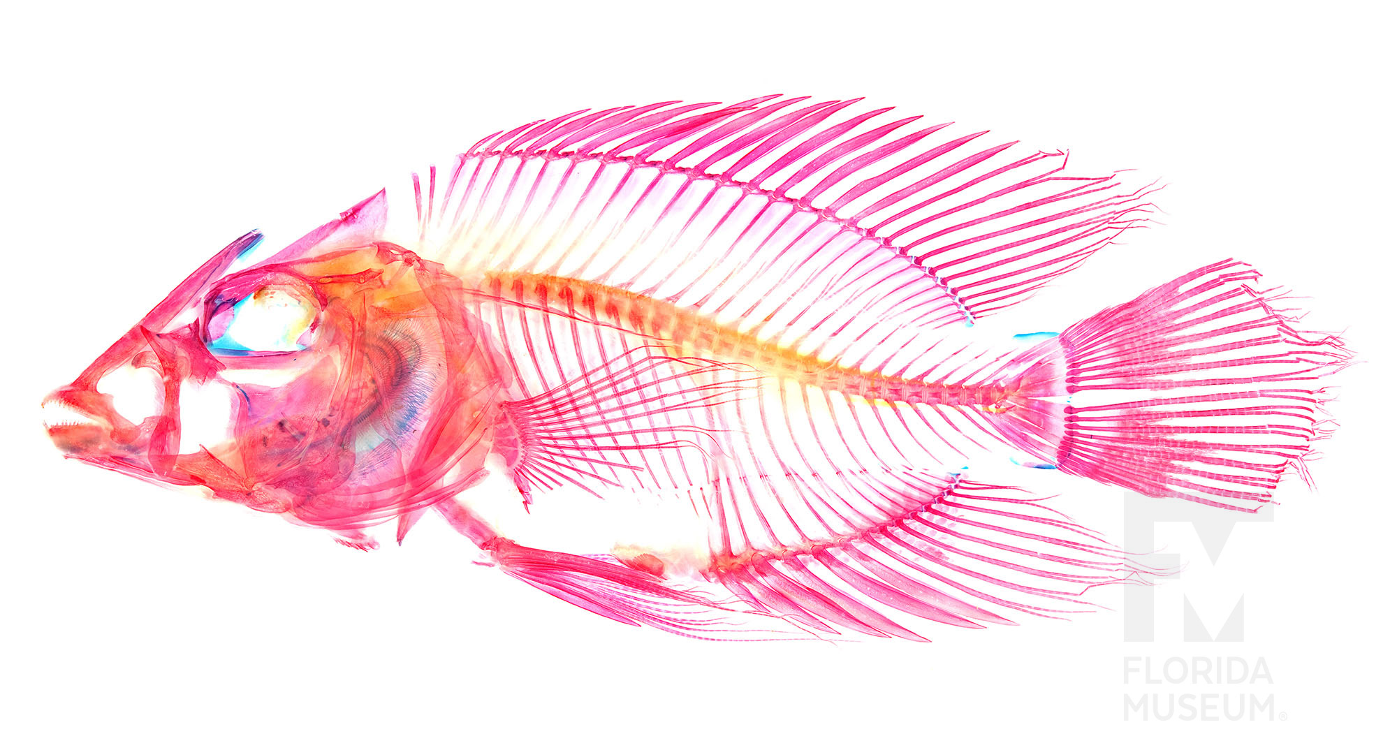
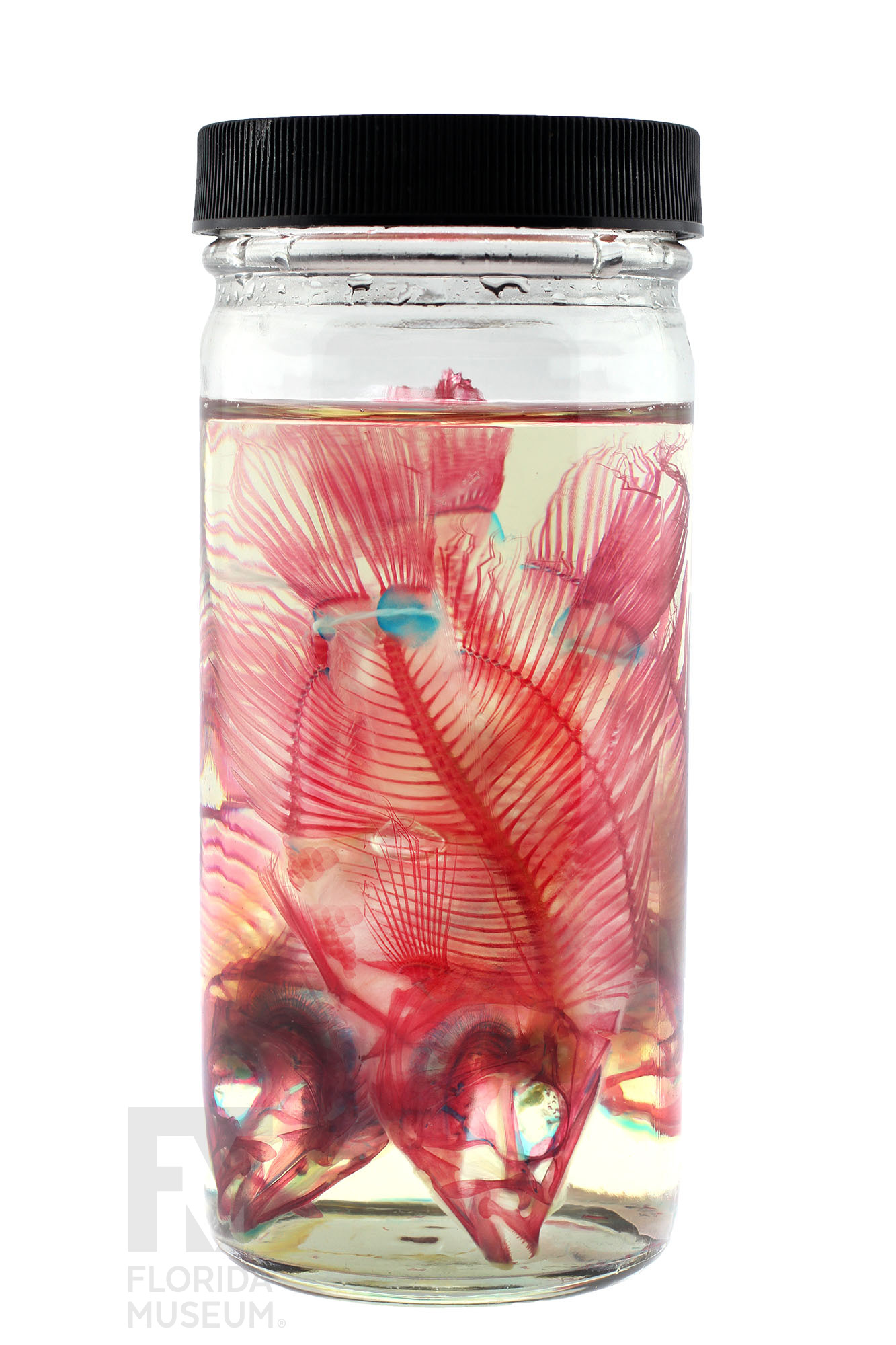
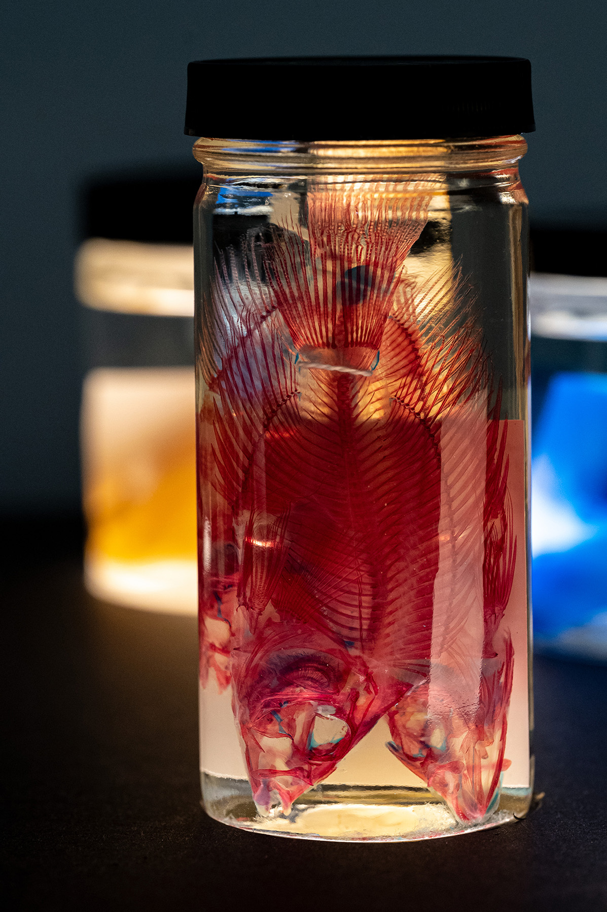
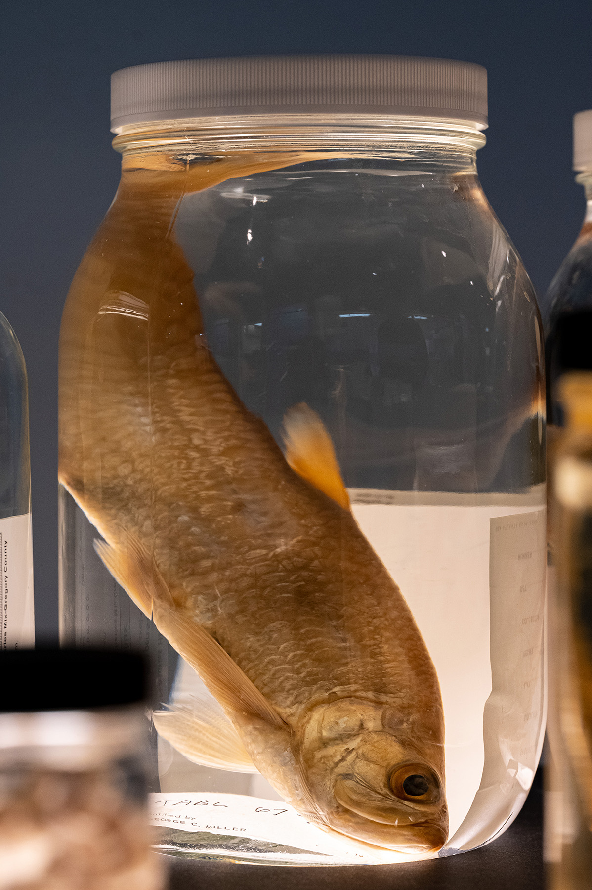
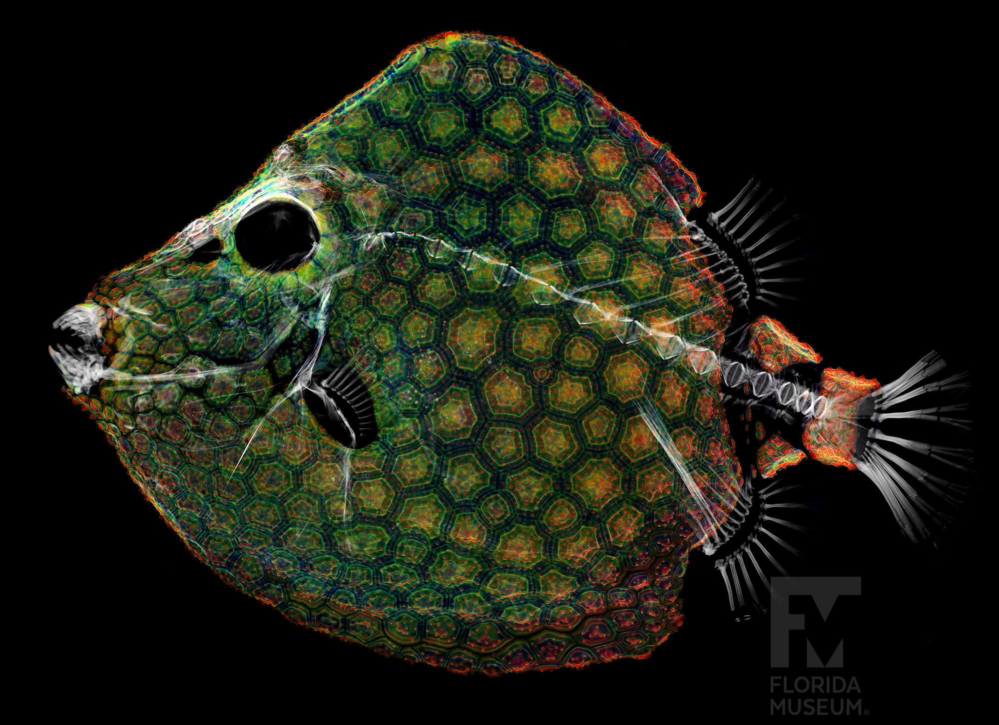
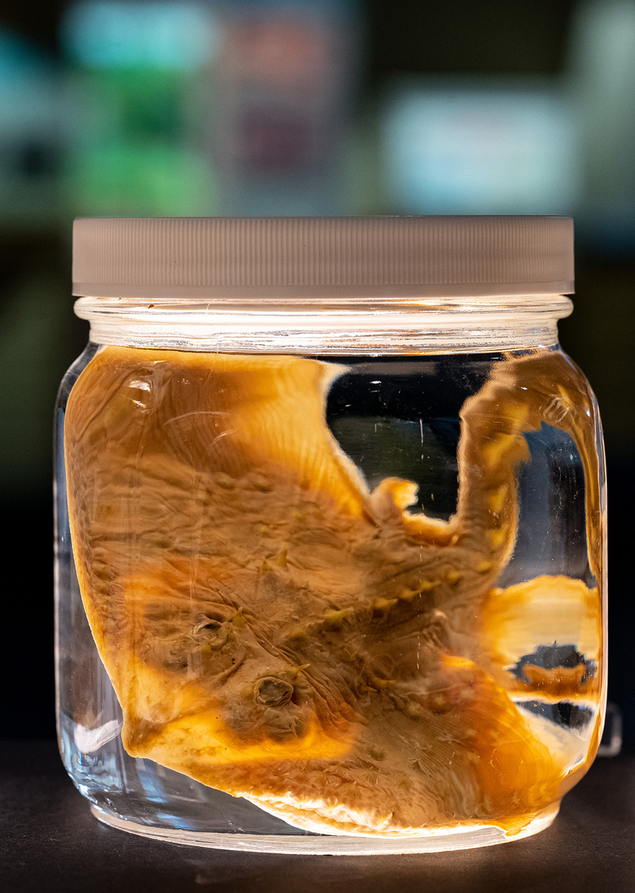
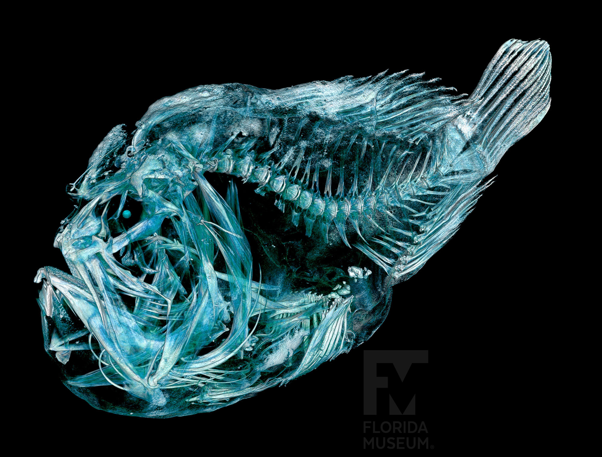
This exhibit showcases specimens from the Florida Museum’s fish collection revealing the beauty in the process of clearing and staining (also known as diaphonization). This process uses a digestive enzyme called trypsin to clear away soft tissue but not the collagen, which supports the skeleton in place. Chemicals are used to stain cartilage blue and bone red. This method has been used for over 100 years and is still used today in natural history collections, remaining an affordable and easily accessible option.
The specimen is stored in glycerin which makes the specimen look clear because glycerin has the same refraction index (how light spreads from one medium to another) as the collagen, the material supporting the skeleton.

Watch the process
- Pros: Affordable and easy to do by following a recipe. It’s also popularly used by biologist/anatomists unsure about which structures are made of cartilage and/or bone.
- Cons: The process is non-reversible and the specimen is forever cleared and stained. Glycerin (material the specimen is stored in) is very sticky and gets on everything!
Explore cleared and stained specimens
Scientific name: Gymnura micrura
Native Distribution: Western and eastern Atlantic Ocean and Gulf of Mexico
Habitat: Marine
Specimen: Florida Museum, UF 51071
Learn more: Florida Museum, Smooth Butterfly Ray profile
Scientific name: Merluccius sp
Native Distribution: Atlantic and Pacific Oceans
Habitat: Marine
Specimen: Florida Museum, UF 186892
Learn more: Fishbase.se
Scientific name: Amia calva
Native Distribution: Mostly eastern North America
Habitat: Freshwater
Specimen: Florida Museum, UF 18751
Learn more: Florida Museum, Bowfin profile
Scientific name: Caquetaia kraussii
Native Distribution: Colombia
Habitat: Freshwater
Specimen: Florida Museum, UF 17200
Learn more: Fishbase.se
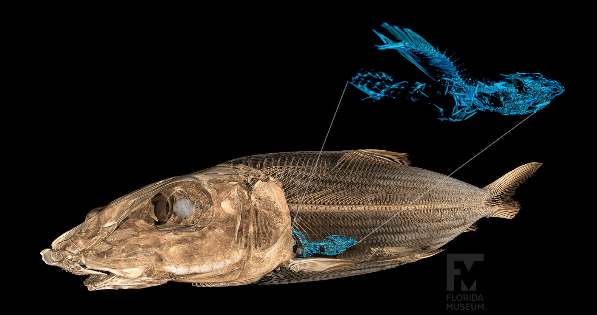
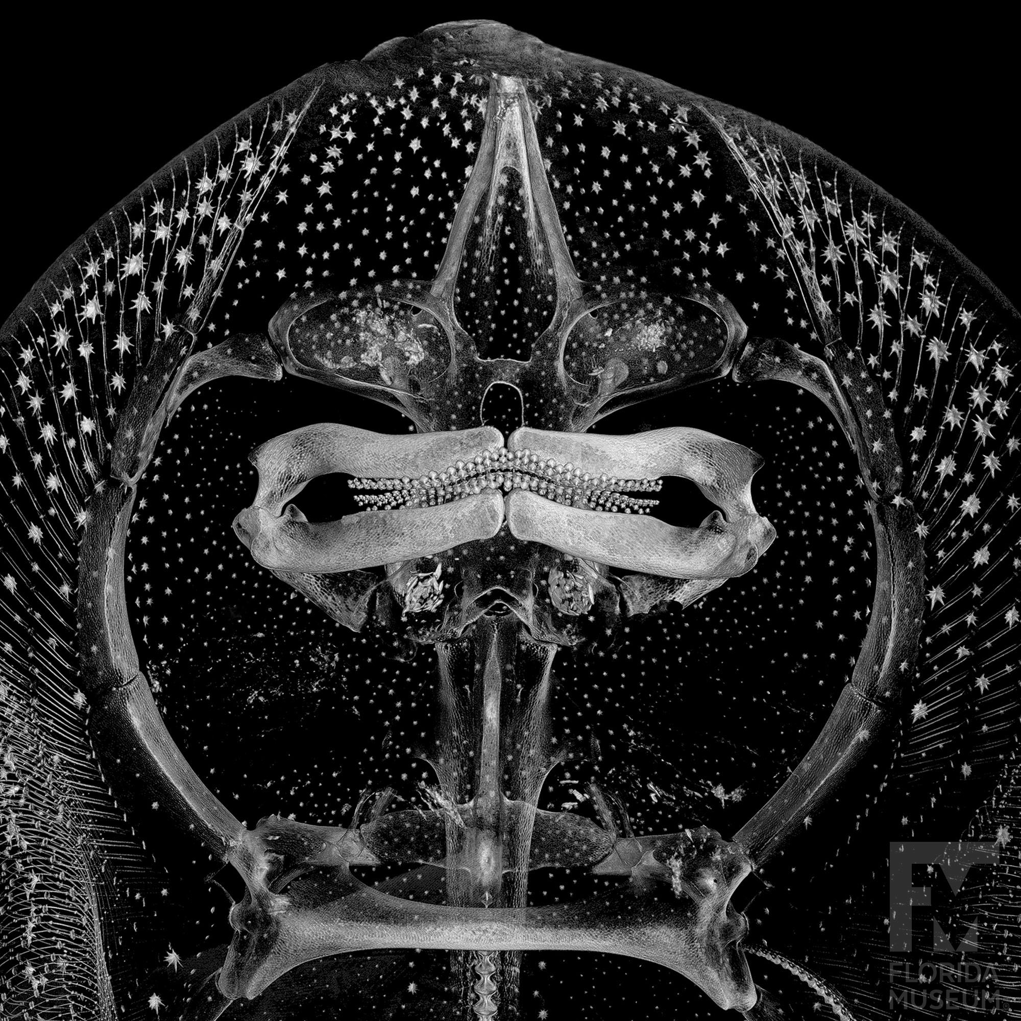
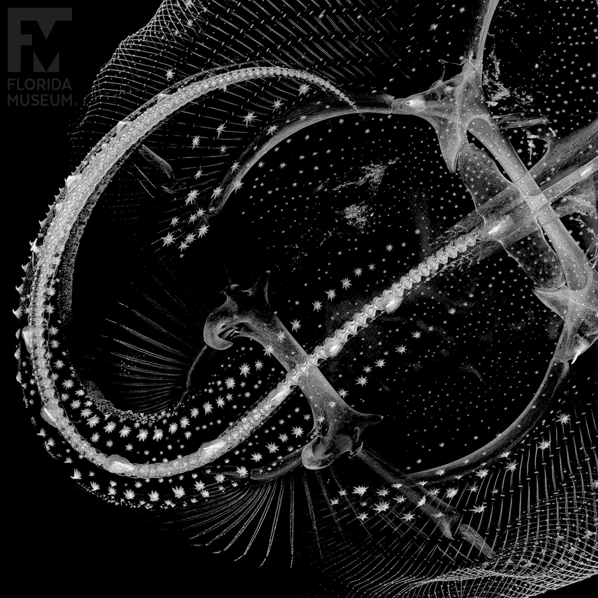
Looking to the Future: CT Scanning for Accessibility & Preservation
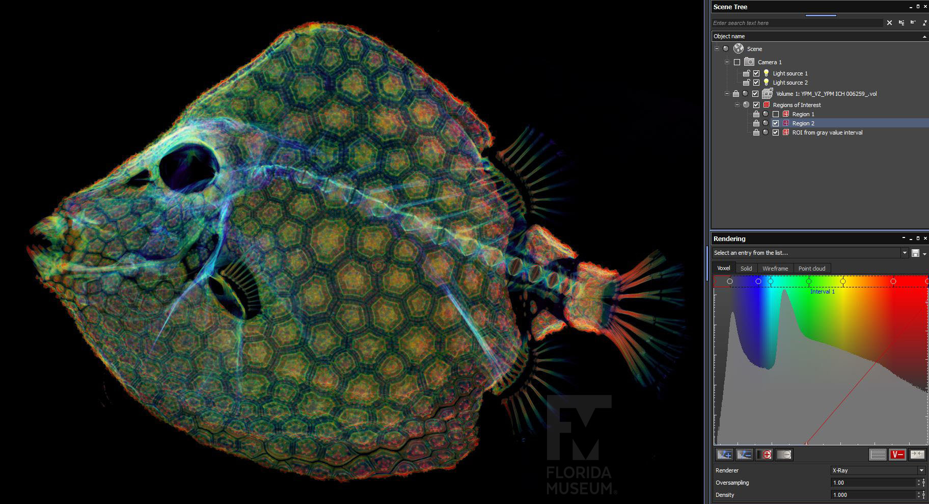
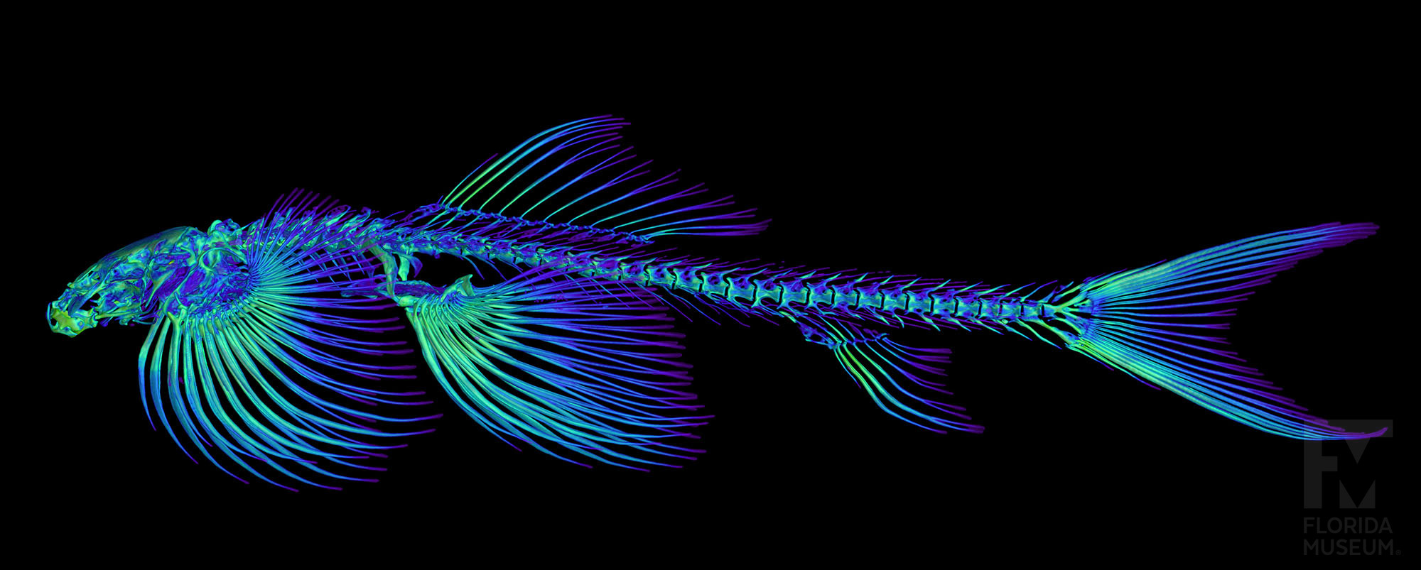
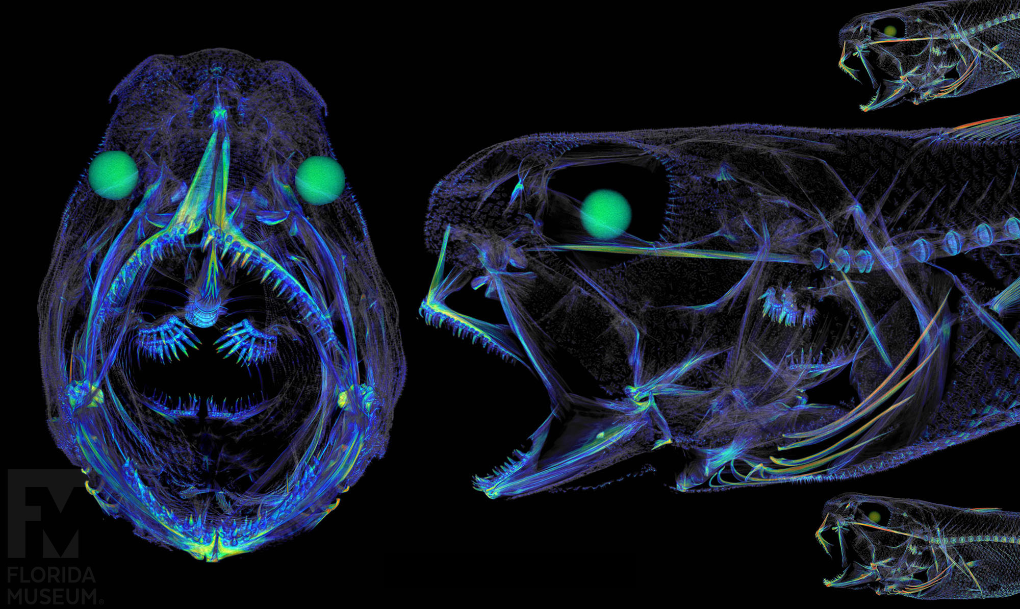
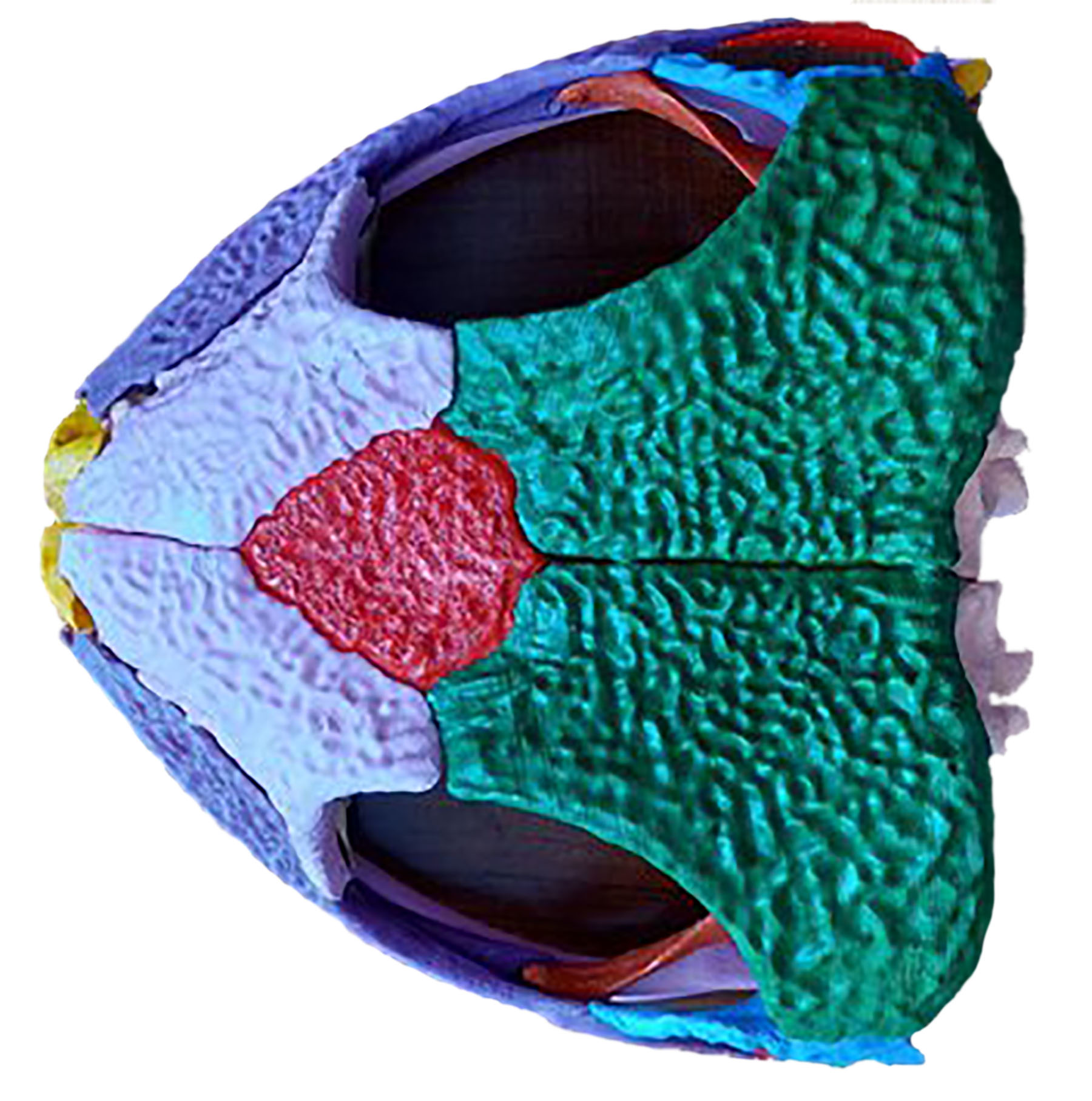
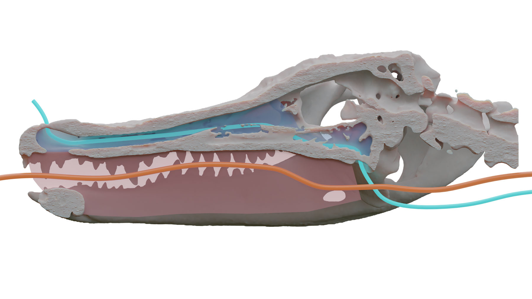
The Florida Museum and other natural history museums are using industrial micro CT scanners to capture a specimen’s anatomy in a non-destructive way. Through the use of x-rays, they digitally capture soft tissue and skeletal anatomy in 3D without causing damage or alteration to the specimen. Similar to getting a CT scan at the hospital, they capture hundreds to thousands of x-ray images of a whole specimen in a 360- degree rotation. These 2D x-ray images are then converted into slices (tomograms) and combined together to make a 3D model (volume). Preserved fish can be kept in these machines for hours without drying out or becoming damaged since they are packaged in plastic bags that help retain moisture.
This process makes the skeleton digitally and globally available for researchers, teachers, students and artists to use. CT data can be rendered different ways. The CT images shown in this exhibit are just one visual interpretation of the scan data.
Watch the process
Pros: Non-destructive and does not require dissection of the specimens. The data can be easily shared and accessed by anyone around the world.
Cons: Scanning can be expensive and not always accessible. CT rendering software can be expensive and powerful computers are needed.
Explore scanned specimens
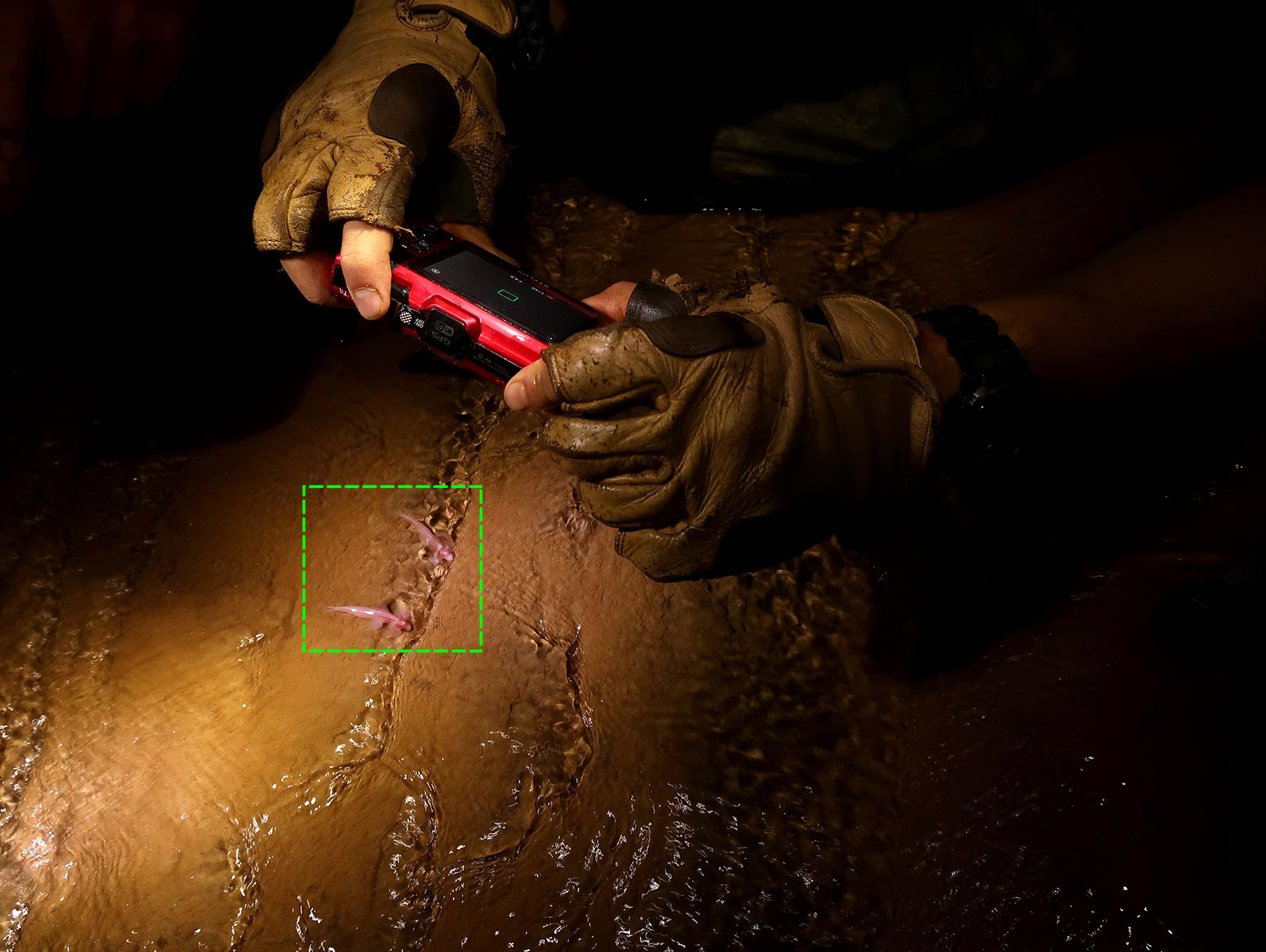
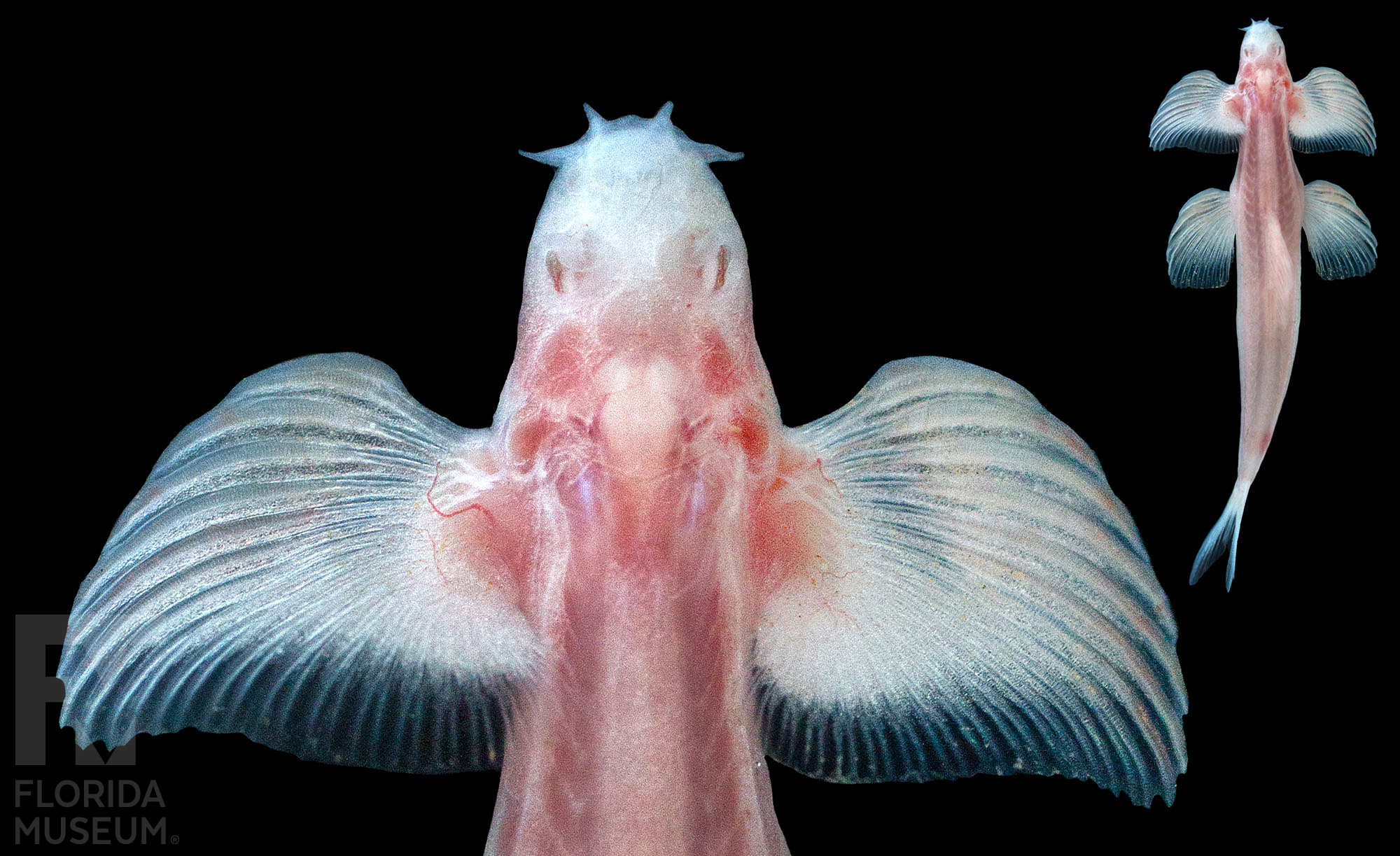
Scientific name: Cryptotora thamicola
Native Distribution: Only found in the caves of northern Thailand
Habitat: Freshwater
Specimen: Scanned on loan from Maejo Aquatic Resources Natural Museum, (MARNM 7413)
Learn more: Fishbase.se
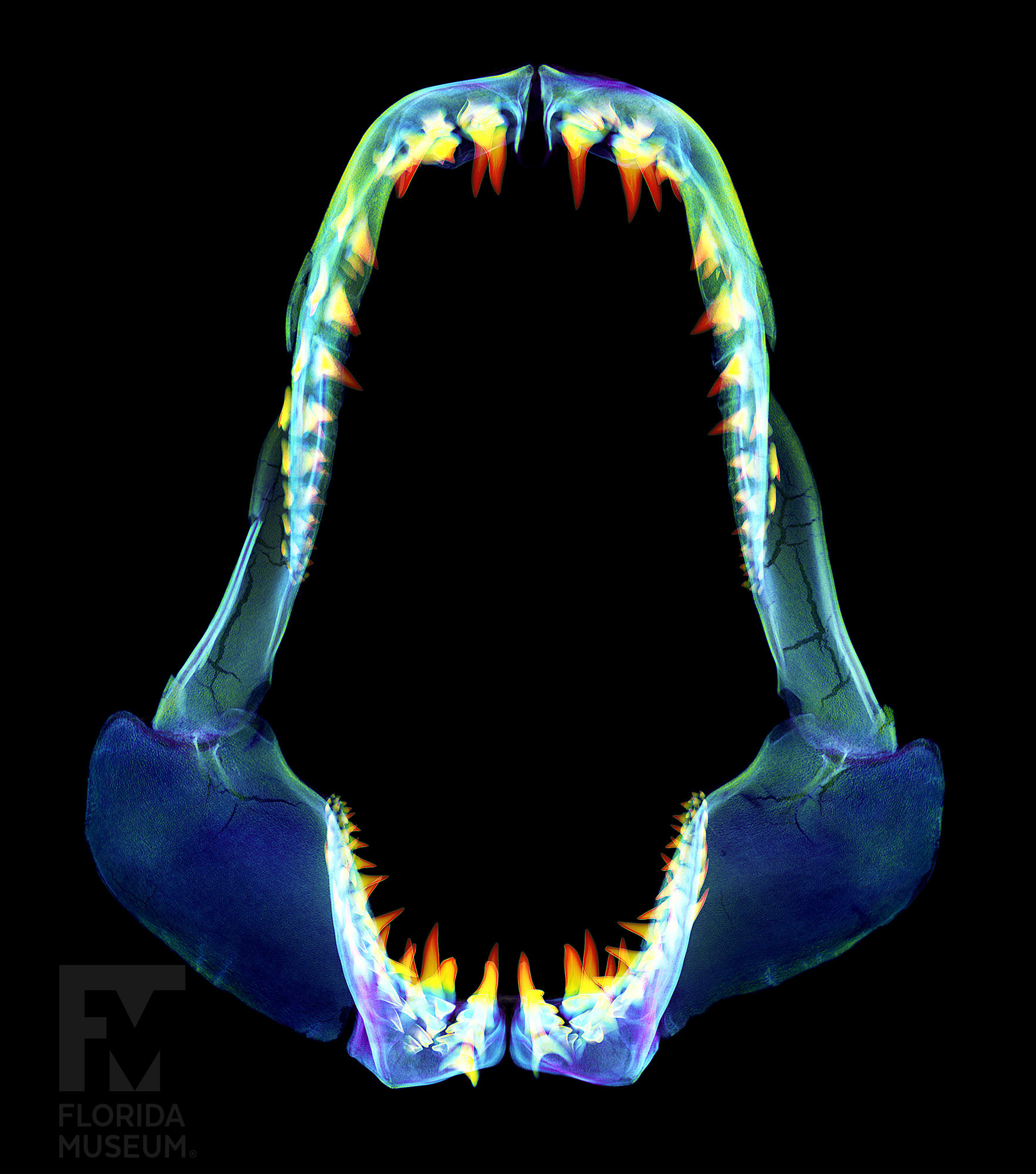
Scientific name: Isurus oxyrinchus
Native Distribution: Worldwide in tropical and temperate waters
Habitat: Marine
Specimen: Florida Museum, UF 242078
Learn more: Florida Museum, Shortfin Mako Shark
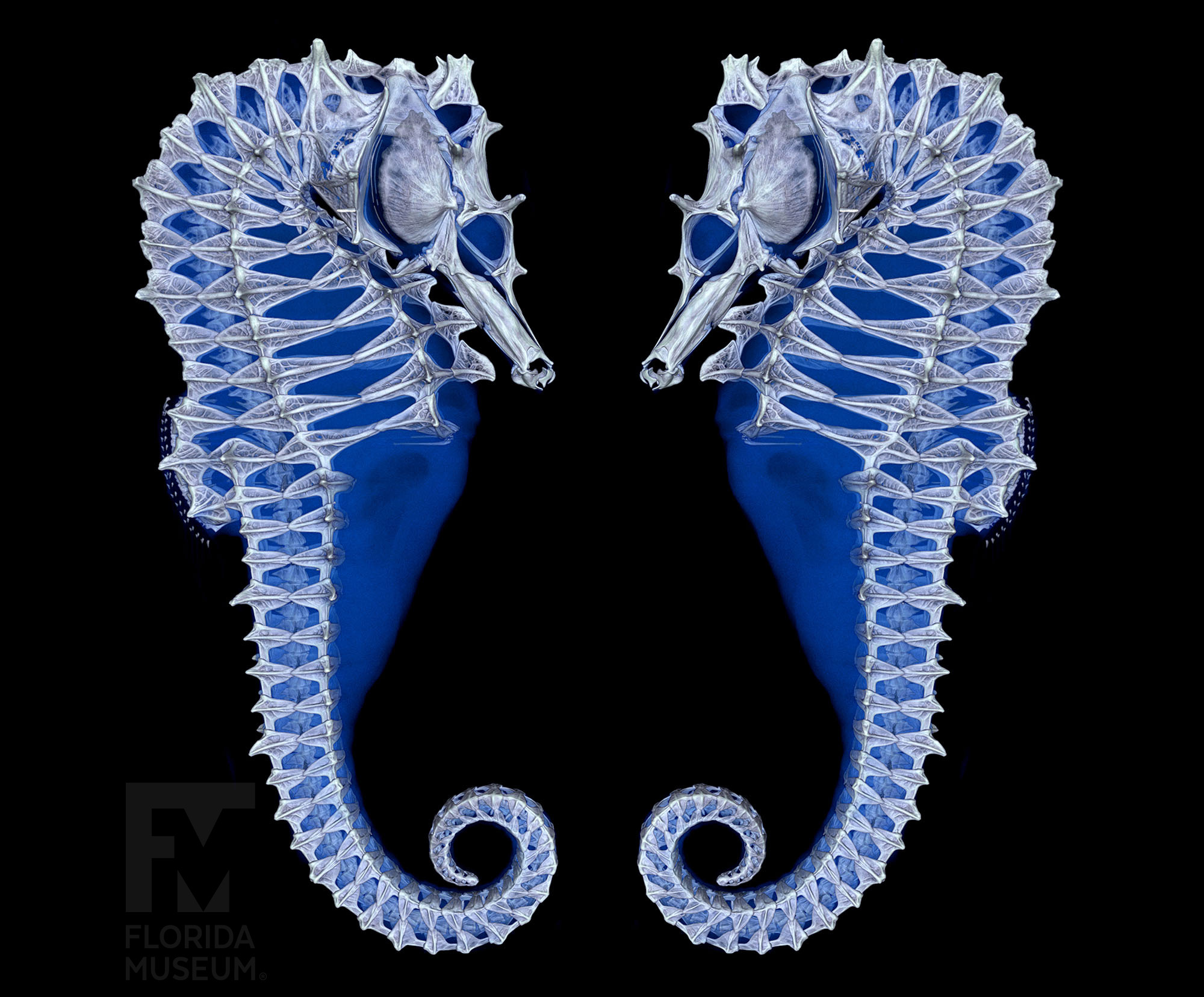
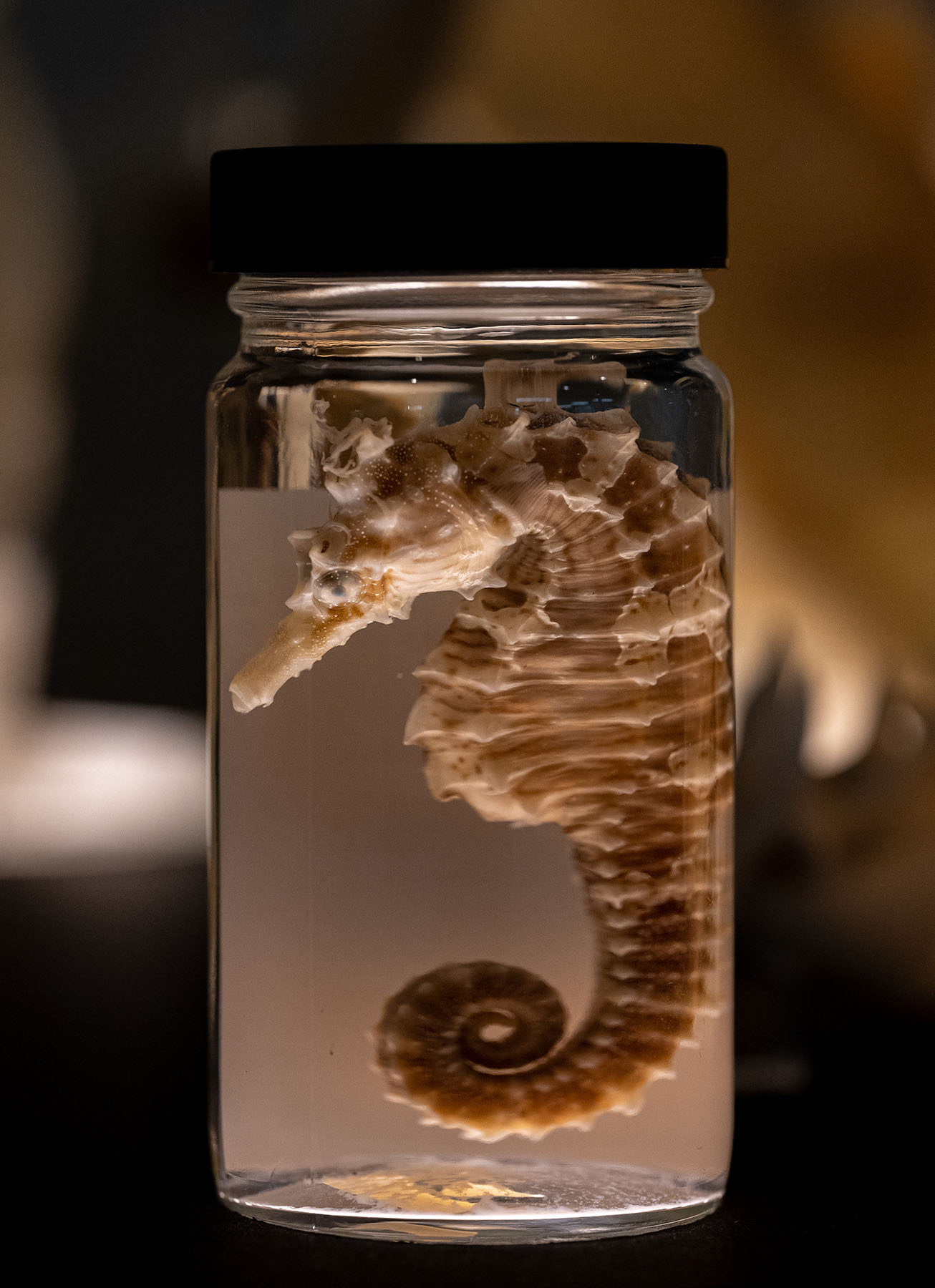
Scientific name: Hippocampus erectus
Native Distribution: From Cape Cod to Argentina and into the Gulf of Mexico
Habitat: Marine
Specimen: Scanned on loan from Texas A&M Biodiversity Research and Teaching Collections, (TCWC 13069-01)
Learn more: Florida Museum, Lined Seahorse
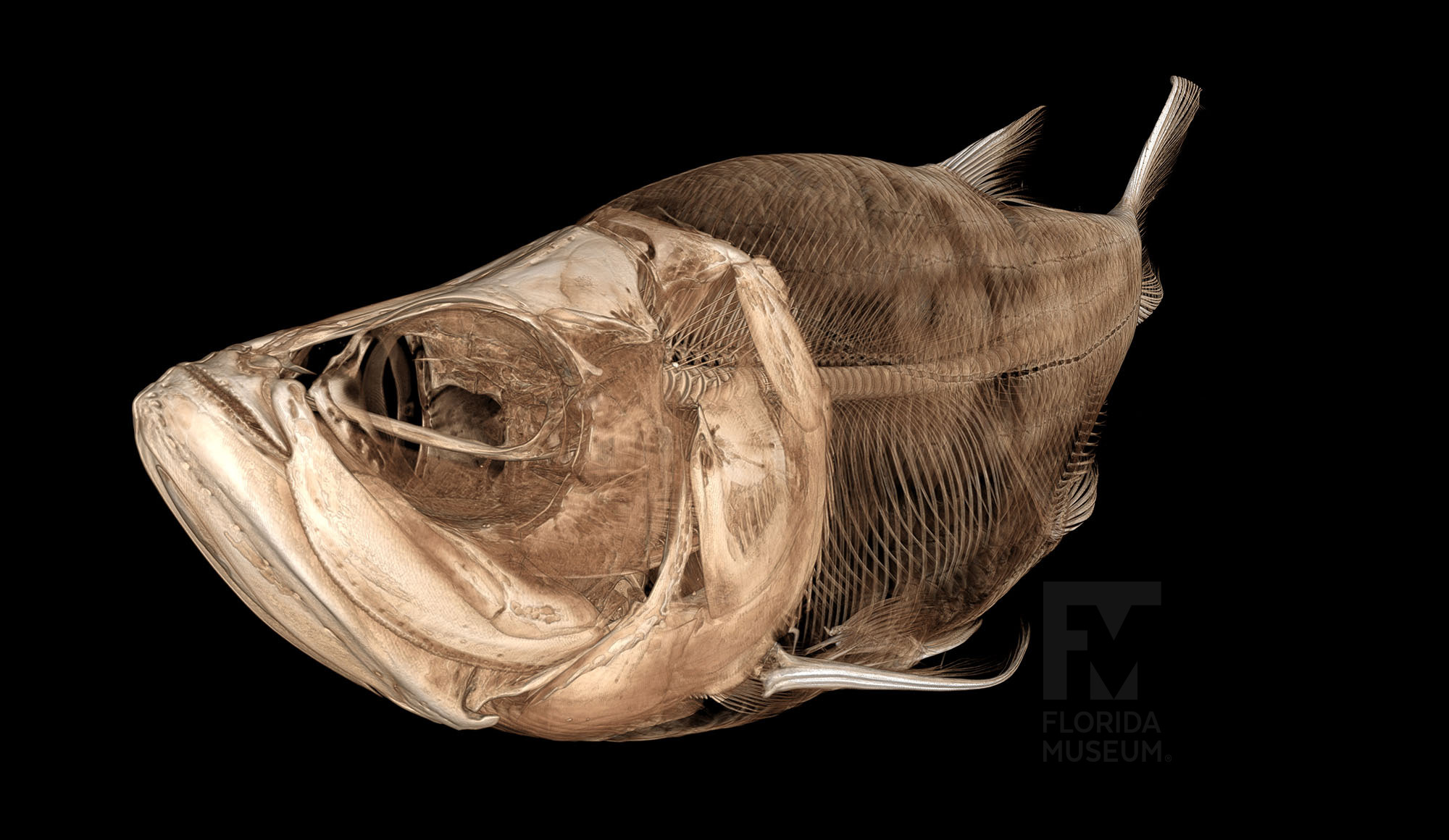
Scientific name: Megalops atlanticus
Native Distribution: Western and eastern Atlantic Ocean and Gulf of Mexico
Habitat: Marine
Specimen: Florida Museum, UF 78923
Learn more: Florida Museum, Tarpon profile
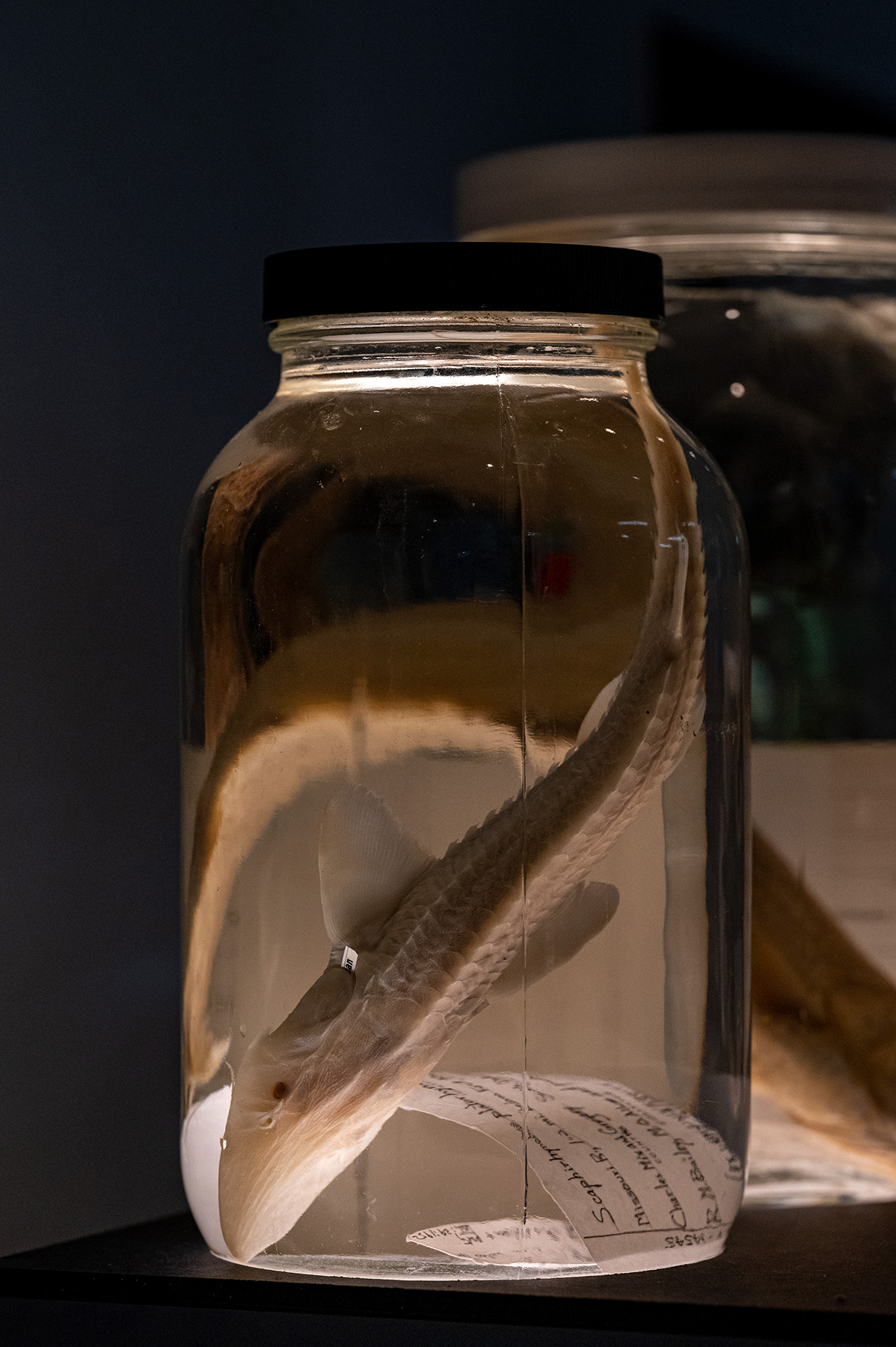
Scientific name: Scaphirhynchus platorynchus
Native Distribution: Mississippi River drainage
Habitat: Freshwater
Specimen: Florida Museum, UF 14545
Learn more: Fishbase.se

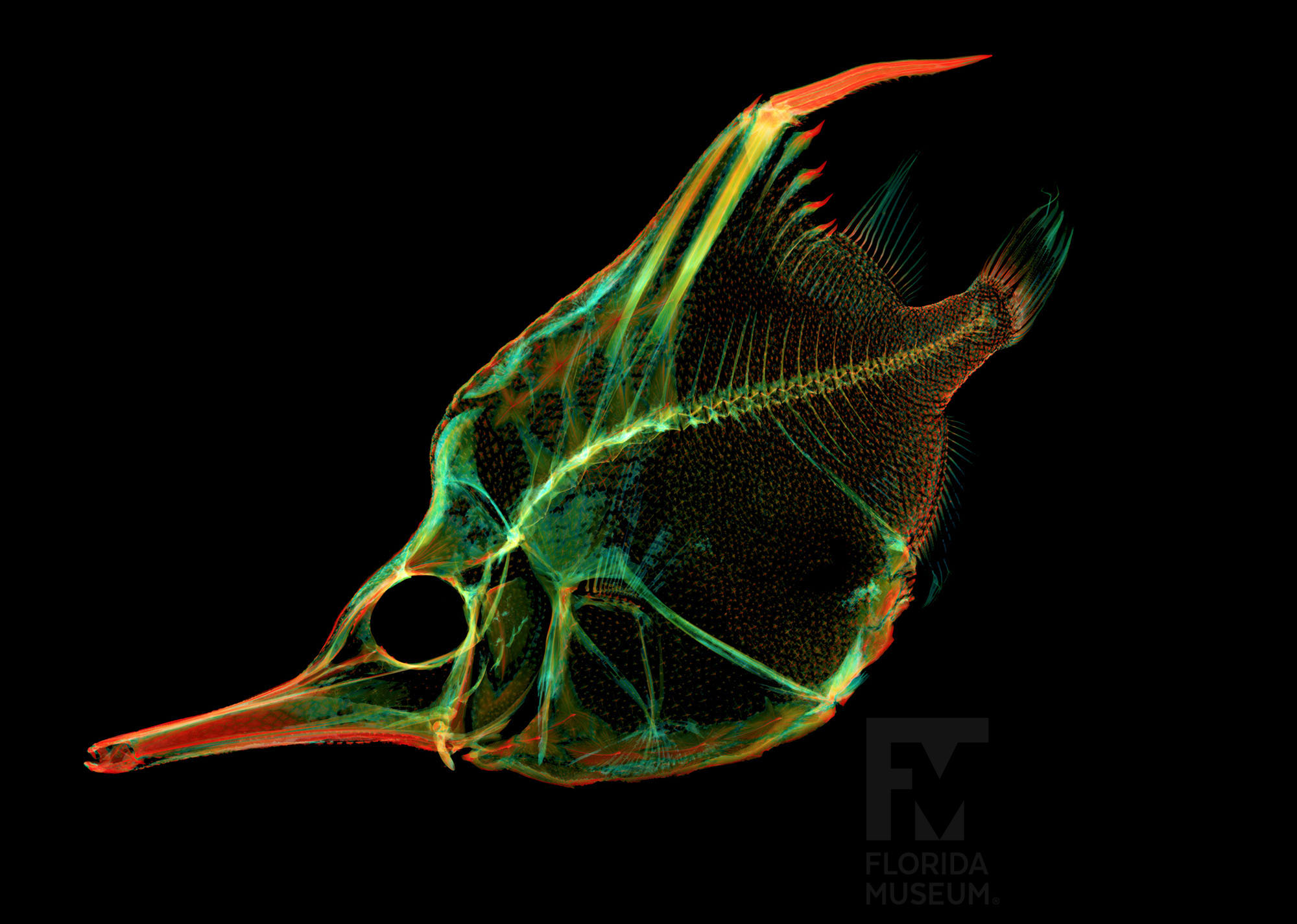
Scientific name: Centriscops humerosus
Native Distribution: Temperate waters in southern hemisphere
Habitat: Marine
Specimen: Scanned on loan from Scripps Institution of Oceanography, SIO 05-134
Learn more: Fishbase.se

Scientific name: Lophius piscatorius
Native Distribution: Native Distribution: Eastern Atlantic Ocean
Habitat: Marine
Specimen: Florida Museum, UF 118531
Learn more: Fishbase.se

Scientific name: Anoplocapros lenticularis
Native Distribution: Eastern Indian Ocean
Habitat: Marine
Specimen: Scanned on loan from Yale Peabody Museum of Natural History, YPM 006259
Learn more: Fishbase.se
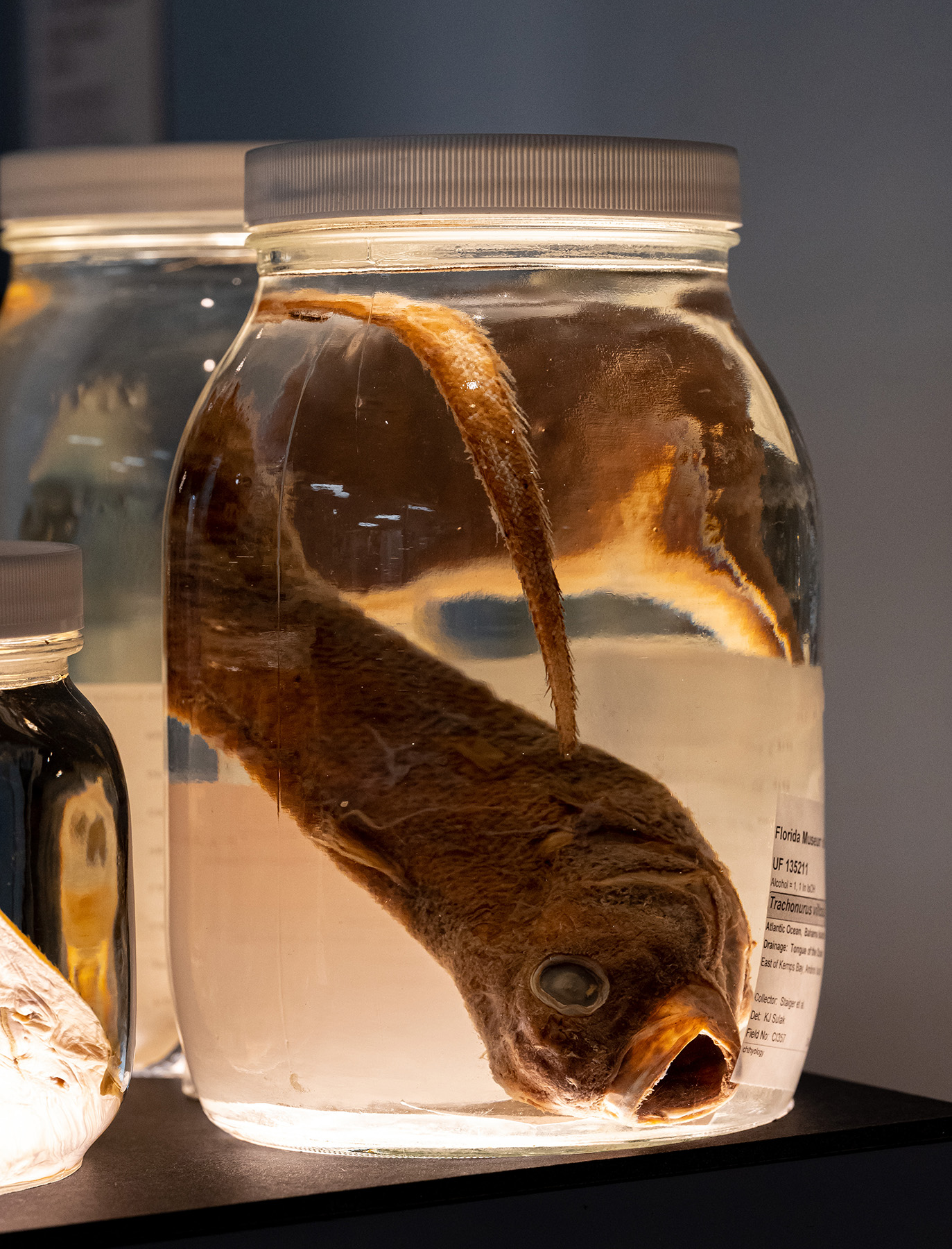
Scientific name: Trachonurus villosus
Native Distribution: Worldwide
Habitat: Marine, deep-water
Specimen: Florida Museum, UF 135211
Learn more: Fishbase.se
Scientific name: Amblyraja radiata
Native Distribution: Eastern and western Atlantic Ocean
Habitat: Marine
Specimen: Scanned on loan from Texas A&M Biodiversity Research and Teaching Collections, TCWC 3220-01
Learn more: Florida Museum, Thorny Skate profile
Research and science communication are changing as global access to digital specimens explode in natural history collections using CT scanner technology.
Museum Specimens at your Fingertips
oVert is a collaborative initiative funded by the National Science Foundation (NSF) that provides free CT data of vertebrates preserved in natural history collections throughout the U.S. All of the data is accessible on MorphoSource, an open-access online image repository. This data provides global digital access to valuable museum collections making them accessible to researchers, educators, students, artists and the public.
oVert-generated media on MorphoSource
- Viewed over a million times
- The oVert project has produced over 10K vertebrate scans
- Downloaded >14,600 times
- Non-research use includes K-12 education and outreach, undergraduate courses and comparative models for artists (3D printing using scan data)
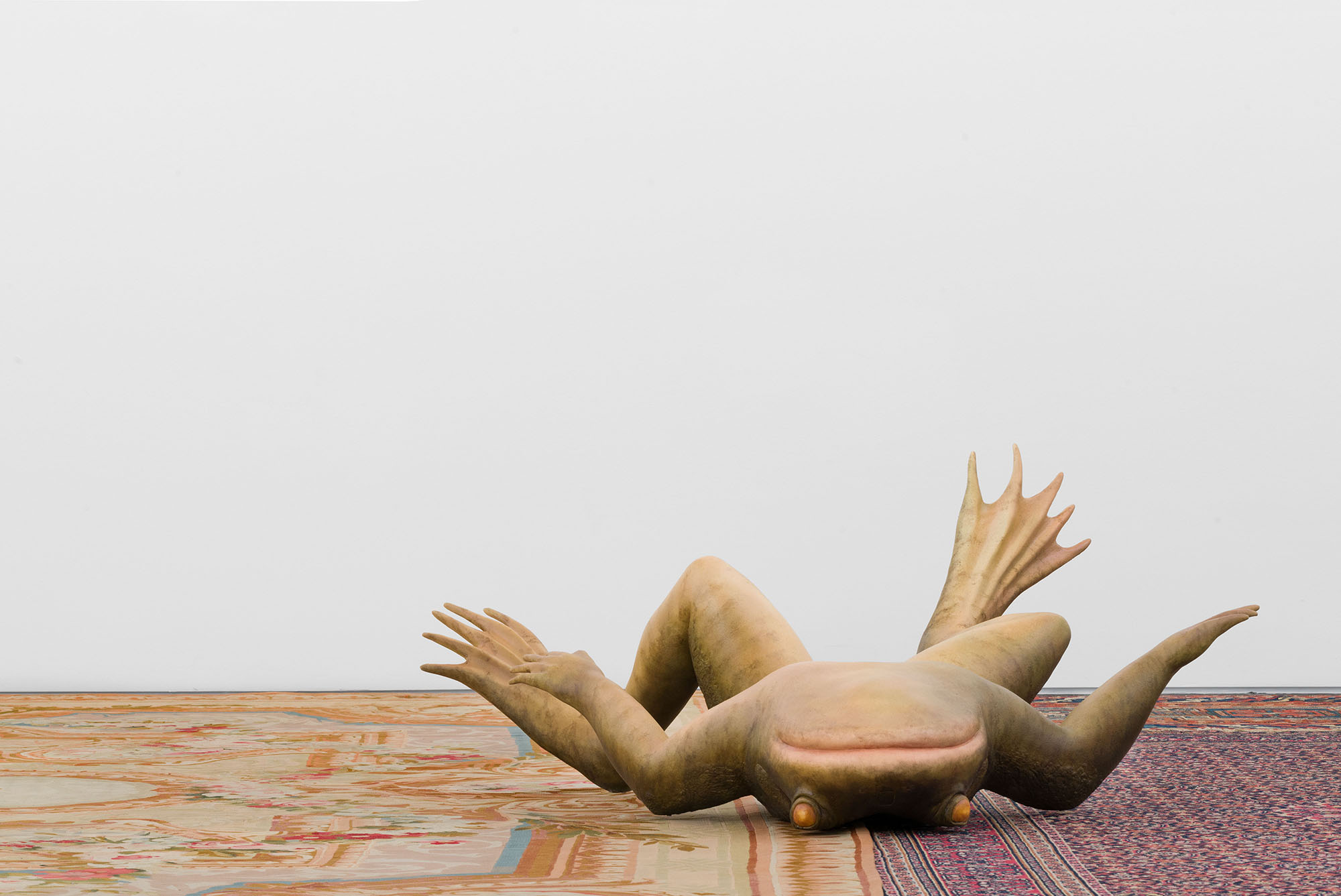
Art & Science
Digital access to natural history specimens is an opportunity for global collaboration and for better understanding and communication of Earth’s biodiversity. Integrating STEAM disciplines (science, technology, engineering, art and math) can make science communication more accessible. Combining visual arts and science may help people relate to an organism’s biology whereas they might get lost in complicated scientific terminology. As new technologies for visualizing data develop, communicating about science will become even more approachable.
 Enlarged printed frog skull identifying every bone of the skull. Photo courtesy of Ramón González Cabrera
- This frog statue in a Carnegie Museum of Art exhibit was created in part from data generated by the oVert project. Photo courtesy of Margaret Honda
- Digital model created from CT data shows the air passageways of an alligator. Photo courtesy of Dominique Adriaens
About Zach
Zachary Randall is an ichthyologist and imaging lab manager at the Florida Museum. He studied photography and freshwater fish systematics during his undergraduate and graduate degrees at UF. He uses visual arts as a form of science communication and utilizes imaging technology to digitize biodiversity collections for research, education and outreach. One of his most memorable moments working at the Florida Museum was the first time he saw a cleared and stained specimen, what seemed like hidden treasure, that he knew he wanted to share.