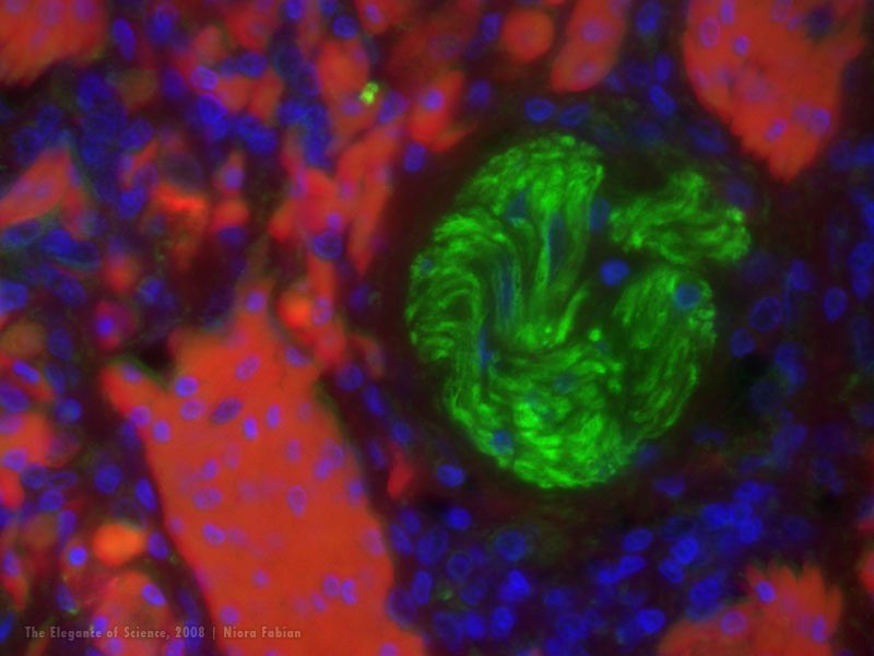
These images were generated in a preliminary part of my graduate thesis research project. I am using immunofluorescence labeling to study tissues of alligators infected with bacteria called Mycoplasma alligatoris. The stains can help visualize individual bacteria within the tissues as well as indicate molecular changes in the cells that reflect an immune response to the pathogen. The tissue samples are collected from necropsy, then processed so that they can be cut in ultra-thin sections (5 microns thick). Then they can be stained for various cellular components of interest and studied under the microscope.