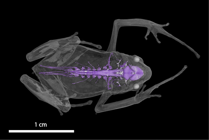
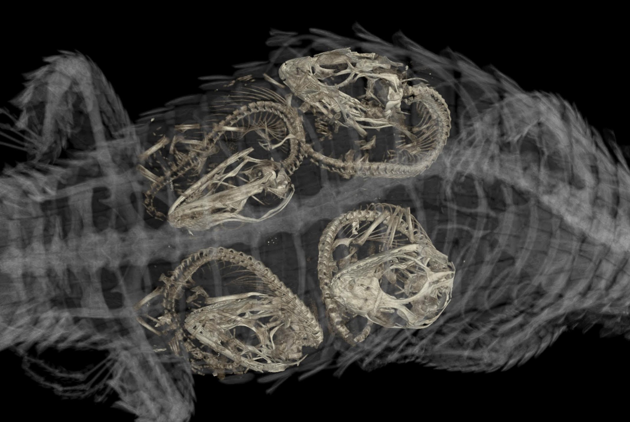
Florida Museum scan by David Blackburn
Encased in hard rock, the bones of many fossilized mammals are only partially visible for scientists to study. A poor attempt to take apart the rock and view the complete fossil may damage the bone, but micro-CT scanning technology has safeguarded the fate of these specimens, many of which are tens of millions of years old.
“You can essentially cut into the specimens in a non-destructive way with a micro-CT scanner,” said Jonathan Bloch, curator of vertebrate paleontology at the Florida Museum of Natural History. “And it allows you to look at the internal anatomy of fossil skulls.”
The new micro-CT scanner at the University of Florida’s Nanoscale Research Facility will allow Bloch and other scientists to closely view and study specimens as small as a micron (one millionth of a meter).
Holding a tiny Notharctus skull in his hand, Bloch explained the scanner can digitally recreate the 45-million-year-old lemur-like primate’s brain, which could give insight into how its body structure differs from modern primates.
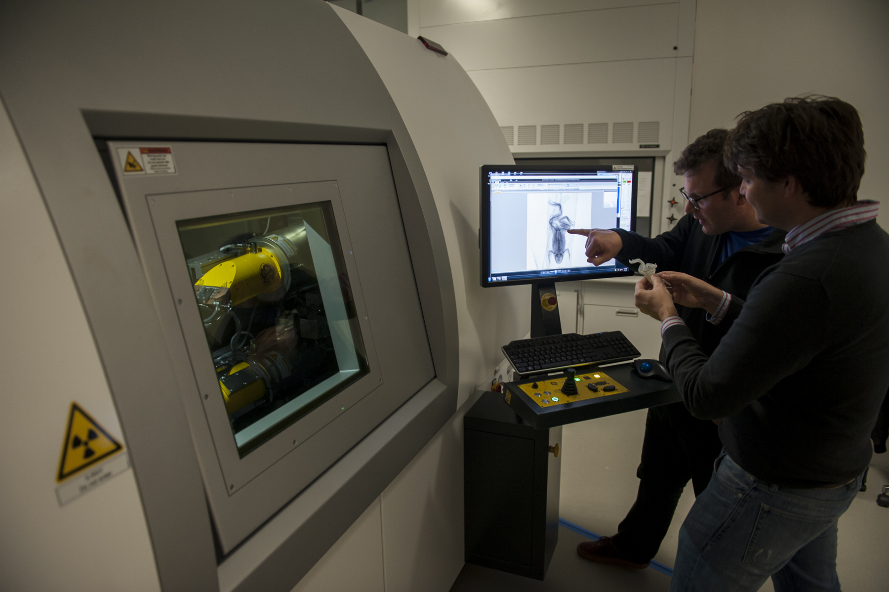
Florida Museum photo by Kristen Grace
“Once you have an image of the brain, you can follow nerves and blood vessels from the brain through the rest of the skull to determine where they go,” he said. “It allows us to test ideas about what these holes and grooves and things are on the surface of fossil bones.”
Florida Museum Associate Curator of Herpetology David Blackburn plans to use the scanner to merge together what looks like a series of puzzle pieces—fossilized frog bones from 25 million years ago—which he keeps ready for scanning in a small box in his office.
“These are parts that we think are all from the same species,” Blackburn said, holding up a bone about 1 centimeter long. “We don’t have a complete one, but we can virtually put it back together.”
“It’s such a great way of sharing our research,” Blackburn said. “We can virtually take specimens apart. We can zoom into any piece of it, we can go inside pieces of it.”
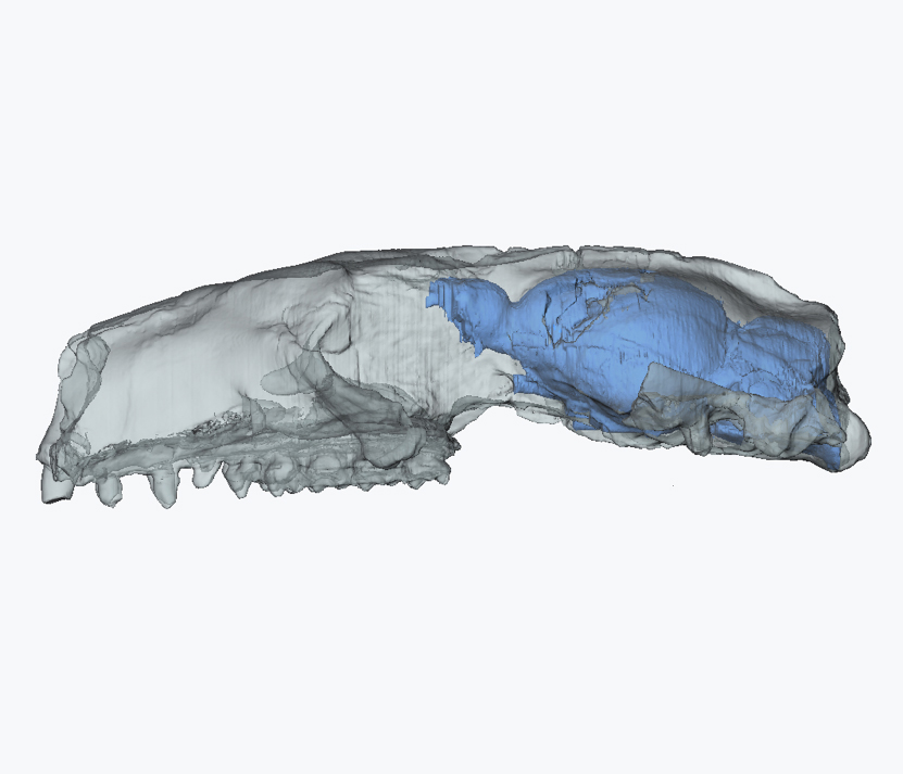
Florida Museum scan by Jonathan Bloch
The scanner works a lot like a traditional X-ray machine one might use at a doctor’s office, but instead of taking an image from one direction, thousands of images are taken from different angles so they can be combined to produce a 3-D picture, Blackburn said.
Bones become visible in X-ray imaging because they are the densest part of an animal’s body, but some of Bloch’s work has involved “filling” empty spaces between bones to outline the size and shape of internal organs. The process is significant in understanding differences between the internal development of fossilized animals from millions of years ago and modern species—it enables scientists to make accurate physical comparisons and unlock their historic connections.
“For the first time, we’re allowed to segment out the shape of the brain inside the skull of these fossil animals without removing the rock or the bone itself,” Bloch said.
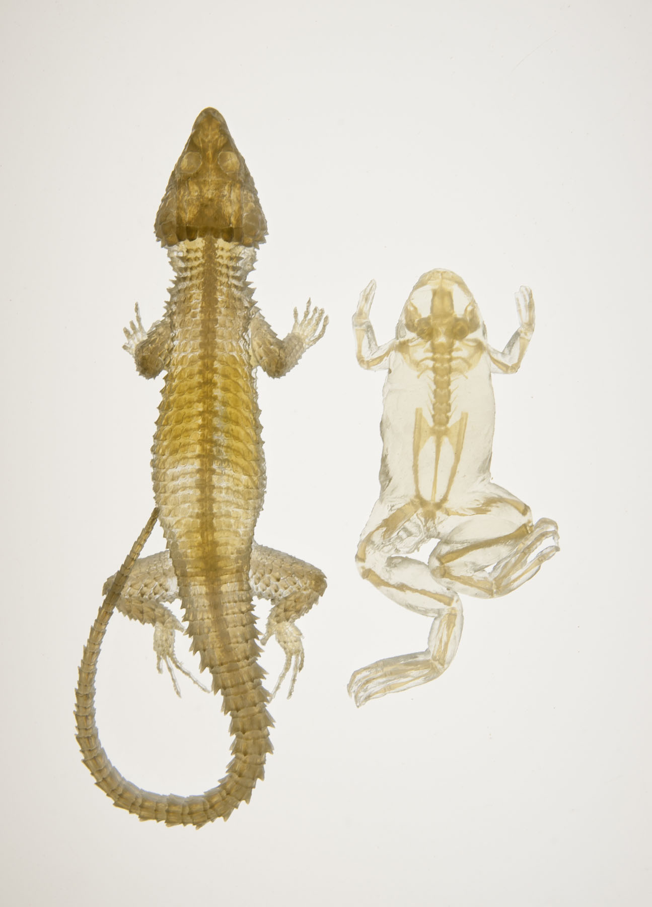
Another way to view internal anatomies with micro-CT scanning involves infusing a specimen with a chemical like iodine that makes soft tissues appear dense while still intact inside the animal—everything from muscles to nerves and blood vessels.
“We can reconstruct a frog’s muscles, see its nervous system, all of its internal organs,” Blackburn said. “Some of this is useful for describing new species, some is simply of interest to understanding basic biology.”
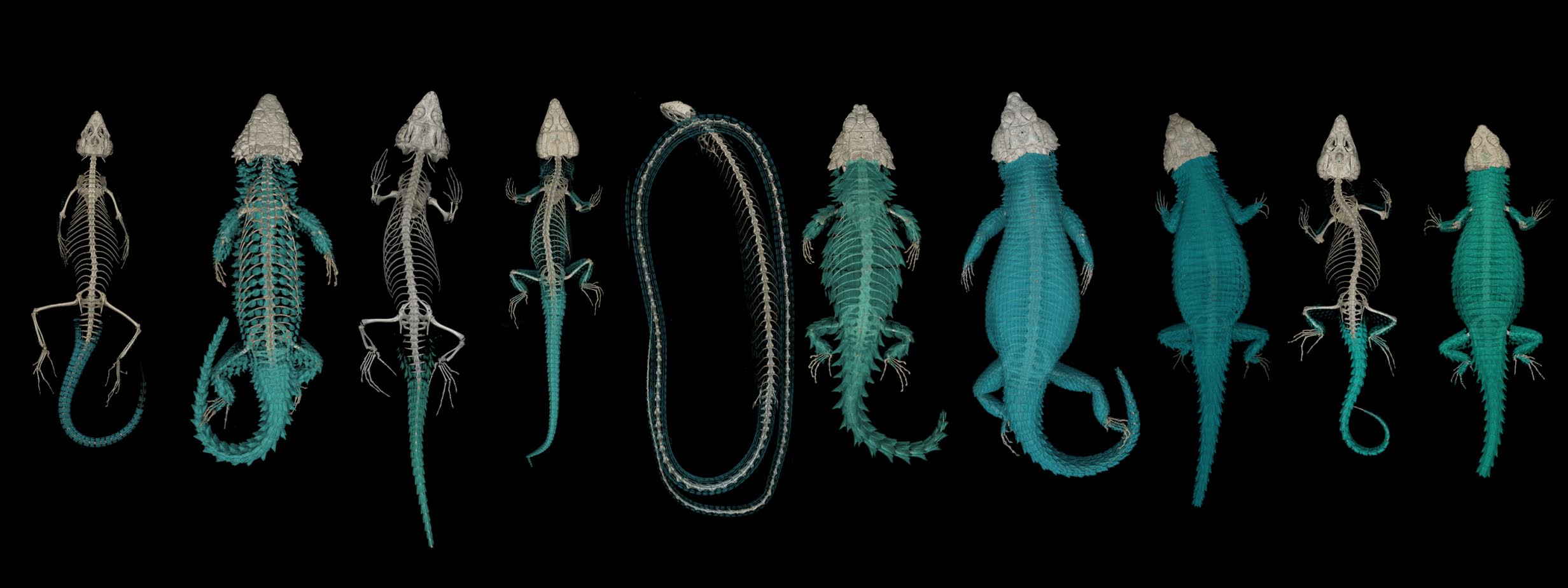
Florida Museum scan by Edward Stanley
The new technology also allows scientists to study some of the smallest and most delicate biodiversity, such as the moth eyes and antennae studied by Akito Kawahara, an assistant curator at the Florida Museum’s McGuire Center for Lepidoptera and Biodiversity.
“When you are working with specimens this small, pieces might get lost,” Kawahara said. “Our research with micro-CT scanning will help us understand how these eyes and antennae vary without dissecting their structure.”
Kawahara said the technology will also help visitors better understand the organisms displayed at the museum.
“To be able to do that without harming any of these really small insects is a great benefit,” he said.
Florida Museum scan by Edward Stanley
The technology may also benefit students in the classroom and members of the public through printed 3-D models of specimens, including the ancient tuatara lizard that is a sister to nearly all living lizard species. Blackburn has a 6-inch tuatara model in his office—about 10 times the size of the original species.
“One plan is to use the technology for generating data for classrooms,” Blackburn said. “We might bring the real specimen into an undergraduate class to show it in a lab, but we’re certainly not going to let a 5-year-old handle it during a public program. A model can be replaced.”
Florida Museum photo by Kristen Grace
A 3-D specimen model will also allow scientists to complete statistical analyses and more accurately measure the size and shape of different body parts. This capability is important for Blackburn and Bloch, who often try to link the ancestries of fossils in the museum collections with living species.
Micro-CT technology has helped UF assistant anthropology professor Valerie DeLeon and her students research the development of primates and the use of mice as models for human biological behaviors. Like Blackburn, she says access to 3-D specimen models will help students better connect with science.
“A high school science teacher in Alaska could download these 3-D compatible files and print the models to use in their classroom,” DeLeon said. “And it’s all based on having the CT data to start with.”
“One of the really valuable aspects of the micro-CT scanner is it not only creates data for UF researchers to study right here and right now, but it creates this resource of data that will be available way beyond the borders of the university and have a lot of benefit to the research community as a whole,” DeLeon said.
Learn more about the Vertebrate Paleontology Collection and the Herpetology Collection at the Florida Museum.